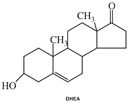
DHEA
Common Names: DHEA, prasterone; DHEA-S, Dehydroepiandrosterone-sulfatePlease read the disclaimer concerning the intent
and limitations of the information provided here.
The information presented in Interactions is for
informational and educational purposes only. It is based on scientific
studies (human, animal, or in vitro), clinical experience, case
reports, and/or traditional usage with sources as cited in each
topic. The results reported may not necessarily occur in all
individuals and different individuals with the same medical conditions
with the same symptoms will often require differing treatments. For
many of the conditions discussed, treatment with conventional medical
therapies, including prescription drugs or over-the-counter
medications, is also available. Consult your physician, an
appropriately trained healthcare practitioner, and/or pharmacist for
any health concern or medical problem before using any herbal products
or nutritional supplements or before making any changes in prescribed
medications and/or before attempting to independently treat a medical
condition using supplements, herbs, remedies, or other forms of
self-care.
![]()
Do not rely solely on the information in this article.
References
Araneo BA, et al. Administration of dehydroepiandrosterone to burned nice preserves normal immunologic competence.
Arch Surg 1993:128:318-325.
Aranen B, Daynes R. Dehydroepiandrosterone functions as more than an antiglucocorticoid in preserving immunocompetetence after thermal injury.
Endocr 1995:136:393-401.
Araneo B, Dowell T, Woods ML, et al. DHEA-S as an effective vaccine adjuvant in elderly humans. Proof-of-principle studies. Ann
NY Acad Sci 1995;774:232-248.
Abstract: Mice immunized shortly after oral dosing with DHEA-S demonstrated high serum antibody titers and complete protection from paralysis. These results became the basis for initiating proof-of-principle studies in human volunteers above age 65 using a licensed influenza vaccine and tetanus toxoid in two independent studies. The use of an oral delivery form of DHEA-S before influenza vaccination was associated with a demonstrable increase in the number of individuals with a fourfold increase in HAI titers following vaccination. The overall mean increase in HAI titers was highest in the DHEAS-treated group. The use of DHEA-S in the immunization of elderly subjects against tetanus toxoid, while unable to enhance the responses, was not a detriment to antibody response.
Arguelles AB, Poggi UL, Saborida C, Hoffman C, Chekherdemian M, et al . Endocrine profiles and breast cancer.
Lancet 1973:1:165-168.
Barrett-Connor B, Khaw K-T, Yen SSC. A prospective study of dehydroepiandrosterone sulfate. mortality, and cardiovascular disease.
N Engl J Med 1986.315:1519-1524.
Barrett-Connor B, Khaw K-T. Absence of an inverse relation of dehvdroepiandrosterone sulfate with cardiovascular mortality in postmenopausal women.
N Engl J Med 1987:317:711.
Barry NN, McGuire JL, van Vollenhoven RF. Dehydroepiandrosterone in systemic lupus erythematosus: relationship between dosage, serum levels, and clinical response.
J Rheumatol 1998 Dec;25(12):2352-2356.
Abstract: OBJECTIVE: To examine in women with systemic lupus eythematosus (SLE) who participated in a clinical trial the relationship between daily dose of dehydroepiandrosterone (DHEA), serum levels of DHEA and DHEA sulfate (DHEAS), clinical effectiveness, and side effects. METHODS: Twenty-three women with mild to moderate SLE were treated with DHEA for a 6 month period. The starting dose was 50 mg/day, and monthly stepwise increases were allowed. Subjects were assessed monthly by the Systemic Lupus Erythematosus Disease Activity Index, Systemic Lupus Activity Measure (SLAM), Health Assessment Questionnaire, and other outcomes. Serum testosterone, DHEA, and DHEAS levels were obtained and side effects noted monthly. RESULTS: Statistically significant improvements were found in all lupus outcomes over 6 months. Serum DHEA and DHEAS levels correlated with the dose of DHEA. Serum DHEA and DHEAS correlated negatively with SLAM score. A second order regression analysis of serum DHEAS level versus SLAM score suggested that the optimal serum level of DHEAS was 1000 microg/dl. The most common side effect was acne. CONCLUSION: The clinical response to DHEA was not clearly dose dependent. Serum levels of DHEA and DHEAS correlated only weakly with lupus outcomes, but suggested an optimum serum DHEAS of 1000 microg/dl. Monitoring these serum levels appears to have limited clinical utility.
Blauer KL, et al. Dehvdroepiandrosterone antagonizes the suppressive effects of dexamethasone on lymphocyte proliferation.
Endocr 1991:129:3174-3179.
Brody S, et al. Adrenal steroids in post-menopausal women: relatiotin to obesity and to bone mineral content.
Maturitas 1987:9:25-32.
Buster JE, Casson PR, Straughn AB, Dale D, Umstot ES, Chiamori N, Abraham GE. Postmenopausal steroid replacement with micronized dehydroepiandrosterone: preliminary oral bioavailability and dose proportionality studies.
Am J Obstet Gynecol 1992;166:1163-1168.
Abstract: Eight postmenopausal women randomly received either a placebo or 150 or 300 mg of oral micronized dehydroepiandrosterone in a lipid matrix. Serum dehydroepiandrosterone, dehydroepiandrosterone sulfate, testosterone, and estradiol were measured periodically over the 12 hours after each dose. Mean peak steroid concentrations after 150 mg (300 mg) doses were dehydroepiandrosterone 1617 (2639) ng/dl, 7 (11.5)-fold above placebo; dehydroepiandrosterone S 1185 (1688) micrograms/dl, 14 (20)-fold above placebo; and testosterone 183 (311) ng/dl, 4 (7)-fold above placebo. Estradiol concentrations remained less than 20 pg/ml, but androgen concentrations rose by 1 hour and remained elevated through the twelfth hour. Peak androgen concentrations and areas under the curves exhibited proportionality with both doses. A testosterone radioimmunoassay revealed a 300% overestimation for testosterone. Thus after appropriate readjustment maximum testosterone concentrations were observed consistently within physiologic premenopausal ranges after the 150 mg dose. The study concluded that micronized dehydroepiandrosterone may provide a steroidal postmenopausal replacement that is adjunctive to estrogens and worthy of further investigation.
Calabrese VP, Isaacs ER, Regelson W. Dehydroepiandrostemone in multiple sclerosis: positive effects on the fatigue syndrome in a non-randomized study. In Kalimi, M, Regelson, W.
The Biological Role of Dehydroepiandrosterone. de Gruyter. New York. 1990. 95-100.
Casson PR, Elkind-Hirsch KE, Buster JE, Hornsby PJ, Carson SA, Snabes MC. Effect of postmenopausal estrogen replacement on circulating androgens.
Obstet Gynecol 1997 Dec;90(6):995-998.
Abstract: OBJECTIVE: To determine the effect of estrogen replacement therapy (ERT) on serum androgen levels in postmenopausal women. METHODS: We measured serum dehydroepiandrosterone (DHEA), DHEA-sulfate, testosterone, estradiol (E2), LH, FSH, and sex hormone binding globulin in 8:00 AM fasting serum samples from a previous randomized, blinded, placebo-controlled crossover study in which 28 postmenopausal women (27 naturally menopausal) were given 2 mg/day of oral micronized estradiol. The treatment arms were 12 weeks with a 6-week washout. RESULTS: Estrogen replacement therapy raised mean (+/- standard error of the mean [SEM]) serum E2 from 8.7 +/- 1.0 to 117 +/- 18.7 pg/mL (P < .001 from baseline). Concurrently, mean (+/- SEM) DHEA-sulfate fell from 67.3 +/- 9.6 to 52.1 +/- 6.4 micrograms/dL (P < .001), and mean (+/- SEM) testosterone fell from 16.1 +/- 2.4 to 9.4 +/- 1.4 ng/dL (P = .006). Both FSH and LH declined significantly. Sex hormone binding globulin increased by 160% with ERT (P < .001). CONCLUSION: Menopausal ERT decreases serum androgen levels, decreasing DHEA-sulfate and testosterone by 23% and 42%, respectively. Whereas the decline in testosterone is likely due to decreased LH-driven ovarian stromal steroidogenesis, the declining levels of DHEA-sulfate also may imply a direct adrenal effect of estrogen. Bioavailable testosterone likely is reduced even more profoundly because sex hormone binding globulin is increased 160% by estrogen. Thus, menopausal ERT may induce relative ovarian and adrenal androgen deficiency, creating a rationale for concurrent physiologic androgen replacement.
Casson PR, Faquin LC, Stentz FB, Straughn AB, Andersen RN, Abraham GE, Buster JE. Replacement of dehydroepiandrosterone enhances T-lymphocyte insulin binding in postmenopausal women.
Fertil Steril 1995;63:1027-1031.
Abstract: Oral micronized DHEA (50 mg/d) was administered in 3-week treatments to 11 postmenopausal women in a prospective, placebo-controlled, randomized, blinded, crossover trial with an interarm washout. After dose (23 hour) serum DHEA, DHEAS, T, and cortisol levels were measured, as were fasting lipoproteins, oral glucose tolerance tests (OGTT), T-lymphocyte insulin binding and degradation, and urine collagen cross-links. Morphometric changes were determined by hydrostatic weighing. Dehydroepiandrosterone sulfate, DHEA, T, and free T increased up to two times premenopausal levels with treatment. Fasting triglycerides declined; no change in collagen cross-links or morphometric indexes was noted. Oral glucose tolerance test parameters did not change, but both T-lymphocyte insulin binding and degradation increased with DHEA. Fifty milligrams per day of oral DHEA gives suprahysiologic androgen levels; 25 mg/d may be more appropriate. Dehydroepiandrosterone enhanced tissue insulin sensitivity and lowered serum triglycerides. Rationale is provided for postmenopausal replacement therapy with this androgen.
Christieff N, Gharakhanian C, Thobie N, et al. Differences in androgens of HIV positive patients with and without Kaposis sarcoma.
J Clin Pathology 1995;48:513-518.
Cleary MP, Shepherd A, Jenks B. Effect of dehydroepiandrosterone on growth in lean and obese Zucker rats. J Nutr 1984:114:1242-1251.
Coleman DL, Leiter EH, Schwizer RW. Therapeutic effects of dehydroepiandrosterone (DHEA) in diabetic mice.
Diabetes 1982:31:830-833.
Davidson MH, Weeks CE, Lardy H, Maki K, Umporowicz D. Safety and Endocrine Effects of 3-Acetyl-7-Oxo DHEA (7-Keto DHEA).
FASEB Jrnl, Abstracts Part II, 4429 (1998)
Abstract: The present study examined the effects of 7-keto-DHEA on several endocrine and safety parameters in healthy adult men (18-49 years of age). 7-Keto-DHEA (n=18) or placebo (n=6) were administered for 8 weeks. The 7-keto-DHEA dose was increased weekly to a maximum of 100 mg bid during the final 4 weeks of treatment. Levels of the hormones measured did not differ between groups (p>0.10) at baseline or during treatment. End-of-trial levels of dihydrotestosterone, estradiol, cortisol and insulin were not different from baseline values (p>0.10). A small reduction in total testosterone [601 ± 142 (SD) vs. 554 ± 149 ng/dL, p<0.01] and a small, clinically insignificant, increase in free testosterone [27 ± 5 vs. 31 ± 8 pg/mL, p<0.01] were observed among men receiving, 7-keto-DHEA. All hormone levels were within normal limits. This study shows that 7-keto-DHEA is well-tolerated at doses up to 200 mg/d and does not produce clinically important sex hormone changes in healthy men.
Daynes R, Dudley DJ, Araneo BA. Regulation of murine lymphokine production in vivo II. DHEA is a natural enhancer of interleukin 2 synthesis by helper T cells.
Eur J Immunol 20:793-802, 1990
Derksen RH. Dehydroepiandrosterone (DHEA) and systemic lupus erythematosus. Semin Arthritis Rheum 1998 Jun;27(6):335-347. (Review)
Abstract: OBJECTIVE: This study was performed to evaluate in vivo and in vitro data on the effects of the adrenal steroid dehydroepiandrosterone (DHEA) with emphasis on its potential use in the treatment of systemic lupus erythematosus (SLE). METHODS: The literature dealing with DHEA was reviewed. RESULTS: Initially, research on DHEA focused on effects of DHEA in relation to obesity. Over the past decade, research stimulated by associations between the physiological decline in DHEA and aging, cardiovascular disease, changes in metabolism, brain function, and immune senescence have generated insight into the many effects that DHEA or its metabolites may have. In SLE a role for sex hormones in both the etiopathogenesis and disease activity is recognized. In SLE, as in aging, low DHEA levels are frequently found, especially with corticosteroid treatment. CONCLUSIONS: Research data in the elderly, on both hormonal and immunologic effects, suggest that DHEA may become an adjunctive treatment for SLE patients.
Dunn PJ, et al. Dehydroepiandrosterone sulphate concentrations in asthmatic patients: pilot study. NZ Med J 1984:97:805-808.
Dyner TS, et al. An open-label dose-escalation trial of oral dehydroepiandrosterone tolerance and pharmacokinetics in patients with HIV disease.
J AIDS 1993;6:459-465.
Ebeling P, Koivisto VA. Physiological importance of dehydroepiandrosterone. Lancet 1994;343:1479-1481.
Gaby AR. Dehydroepiandrosterone: Biological Effects and Clinical Significance.
Alternative Medicine Review. 1996:1(2);60-69.
Gaby AR. DHEA: The Hormone that Does it All. Holistic Medicine. Spring, 1993: 19-24.
Gaby AR. Preventing and Reversing Osteoporosis. Prima Publishing: Rocklin, CA, 1993.
Gaby AR. Research Review. Nutr Healing Jun 1997: 8.
Gennari R, Alexander JW. Arginine, glutamine, and dehydroepiandrosterone reverse the immunosuppressive effect of prednisone during gut-derived sepsis.
Crit Care Med 1997 Jul;25(7):1207-1214.
Abstract: OBJECTIVE: Corticosteroids are used broadly in clinical practice but may profoundly impair resistance to infections. In contrast, arginine and glutamine are safe and effective immunonutrients that can improve resistance to infection in both animals and humans. This study assessed whether arginine and/or glutamine, with or without dehydroepiandrosterone, a natural endogenous steroid, could reverse the susceptibility to infection caused by prednisone in a burned animal model. DESIGN: Prospective, randomized study. SETTING: A laboratory approved by the American Association for the Accreditation of Laboratory Animal Care at the Shriners Burns Institute, Cincinnati Unit. SUBJECTS: Adult female Balb/c mice, weighing 18 to 22 g. INTERVENTIONS: Animals were prefed an arginine- and/or glutamine- or a glycine-supplemented diet for 14 days. Dehydroepiandrosterone (25 mg/kg/day) and/or prednisone (10 mg/kg/day) were given on days -4 to 0 before animals were given a gavage of 10(9) 111indium-oxine-radiolabeled or -unlabeled Escherichia coli and 20% total body surface area burn injury. Survival rate and the extent of translocation of E. coli were determined. MEASUREMENTS AND MAIN RESULTS: Feeding with diets supplemented with arginine, glutamine, and arginine plus glutamine and treatment with dehydroepiandrosterone reversed the susceptibility to infections caused by prednisone and burn injury. The beneficial effects were mediated by enhanced killing of translocated bacteria and/or by an improved gut barrier function. CONCLUSIONS: Dietary supplementation can reverse the susceptibility to infections caused by prednisone. Both arginine and glutamine as well as dehydroepiandrosterone may be useful therapeutic agents for preventing infections in steroid-treated patients.
Hasheeve, D, Salvato, P, Thompson, C. DHEA: a potential treatment for HIV disease. Intl AIDS Conference Abstract no PB0322, 1994.
Helzlsouer, KJ, et al. Relationship of prediagnostic serum levels of dehydroepiandrosterone and dehydroepiandrosterone sulfate to the risk of developing premenopausal breast cancer.
Cancer Res 1992:52:14.
Jacobson, MA, Fusaro, RE, Galznarini, M, Lang, W. Decreased serum dehydroepiandrosterone is associated with an increased progression of human immunodeficiency virus infection in men with CD4 cell counts of 200-499.
J Infect Dis 1991:164:864-868.
Koo E, Feher KG, Feher T, Fust G. Effect of dehydroepiandrosterone on hereditary angioedema.
Klin Wochenschr 1983;61:715-717.
Abstract: Hereditary angioneurotic edema (HAE) is a complement-related clinical disorder with a deficiency of the C1 esterase inhibitor protein. Eight patients with severe attacks of the disease were treated with the adrenal androgen dehydroepiandrosterone sulphate (DS). Steroid therapy for 3-28 months resulted in dramatic improvement in their clinical state and a moderate increase in the serum concentration of C1 inhibitor. There was a significant increase in the serum level of either unconjugated dehydroepiandrosterone (D) or of DS during treatment.
Labrie F, Belanger A, Simard J, et al. DHEA and peripheral androgen and estrogen formation:
Intracrinology. Ann NY Acad Sci 1995;774:16-28.
Loria, RM. et al. Protection against acute lethal viral infections with the native steroid dehydroepiandrosterone
(DHEA). J Med Viral 1988:26:301-314..
Loviselli A, Pisanu P, Cossu E, et al. Low levels of dehydroepiandrosterone sulfate in adult males with insulin-dependent diabetes mellitus.
Minerva Endocrinol 1994;19:113-119.
Abstract: 805 participants were divided into three groups. In all three groups serum concentrations of DHEAS, 17 OH progesterone (17 OHP), delta 4 androstenedione (A4) and cortisol (F) were measured. In 10 patients from group A and in 9 patients from group B the ACTH test (9.25 mg IM Synacthen) was administered and the same hormonal pattern was measured after 60 min. In group C the same hormonal evaluation was performed 5 +/- 2.8 months after commencement of insulin therapy. The study found that DHEA-S serum concentrations were significantly decreased in group A (median 2.9; range 1.1-5.2 mumol/l) with respect to group B (median 5.7; range 3.0-9.5 mumol/l) (p < 0.0012). However, the serum concentrations of 17 OHP (median 3.9 nm/l; range 2.9-6.9 nm/l and A4 (median 5.2 nm/l; range 1.8-10.2 nm/l) were also significantly reduced, while cortisol levels and the 17 OHP/A4 ratio were comparable to group B. After administration of ACTH, the delta increment in cortisol percentage showed a frank increase (55.1%) in group A with respect to group B (33.1%) (p < 0.01). The rise in DHEA-S showed a lower increase in group A (10.2%) with respect to group B (65.5%) even though not statistically significant, while the other hormones showed an overlap between the two groups. In group C the serum concentrations of hormones before insulin therapy did not show any statistical differences with respect to the values in group B. A second evaluation, which was performed during insulin therapy, showed that only the 17 OHP/A4 ratio tended towards higher values with respect to pretherapy values (1.1 and 0.6 respectively; p = 0.07). In conclusion our data confirm low DHEA-S levels during chronic insulin administration therapy. The underlying mechanism could be a general aspecific reduction in the activity of P 450 C 21 SCC enzymes in contrast with the specific inhibition of 17.20-lyase obtained during insulin bolus. Whether the low serum concentrations of DHEA-S can determine an atherogenic effect of insulin needs further investigation, but the hormone could constitute a new parameter for the follow-up of patients affected by diabetes mellitus.
May M, et al. Protection from glucocorticoid induced thymic involution by
dehydroepiandrosterone. Life Sci 1990:46:1627-1631.
Mayer D, Weber E, Bannasch P. Modulation of liver carcinogenesis by dehydroepiandrosterone. In Kalimi, M, Regelson, W.
The Biological Role of Dehydroepiandrosterone. de Gruyter. New York. 1990. pp. 361-385.
McIntosh MK, Berdanier CD. Influence of dehydroepiandrosterone (DHEA) on the thyroid hormone status of BHE/cdb rats.
J Nutr Biochem 1992:3;194-199.
Merril CR, Harrington MG, Sunderland T. Reduced plasma dehydroepiandrosterone concentrations in HIV infection and Alzheimers disease. In: Kalimi, M, Regelson, W.
The Biological Role of Dehydroepiandrosterone. de Gruyter. New York. 1990. 101-105.
Mitchell LE, et al. Evidence for an association between dehydroepiandrosterone sulfate and nonfatal, premature myocardial infarction in males.
Circulation 1994:89:89-93.
Monroe SE, Menon KMJ. Changes in reproductive hormone secretion during the climacteric and postmenopausal periods.
Clin Obstet Gynecol 1977:20:113-122.
Morales AJ, Nolan JJ, Nelson JC, Yen SS. Effects of replacement dose of dehydroepiandrosterone in men and women of advancing age.
J Clin Endocrinol Metab 1994;78:1360-1367.
Abstract: A randomized placebo-controlled cross-over trial of nightly oral DHEA administration (50 mg) of 6-month duration was conducted to study the effect of a replacement dose of DHEA in 13 men and 17 women, 40-70 years of age. During each treatment period, concentrations of androgens, lipids, apolipoproteins, IGF-I, IGF-binding protein-1 (IGFBP-1), IGFBP-3, insulin sensitivity, percent body fat, libido, and sense of well-being were measured. A subgroup of men (n = 8) and women (n = 5) underwent 24-h sampling at 20-min intervals for GH determinations. DHEA and DS serum levels were restored to those found in young adults within 2 weeks of DHEA replacement and were sustained throughout the 3 months of the study. A 2-fold increase in serum levels of androgens (androstenedione, testosterone, and dihydrotestosterone) was observed in women, with only a small rise in androstenedione in men. There was no change in circulating levels of sex hormone-binding globulin, estrone, or estradiol in either gender. High density lipoprotein levels declined slightly in women, with no other lipid changes noted for either gender. Insulin sensitivity and percent body fat were unaltered. Although mean 24-h GH and IGFBP-3 levels were unchanged, serum IGF-I levels increased significantly, and IGFBP-1 decreased significantly for both genders, suggesting an increased bioavailability of IGF-I to target tissues. This was associated with a remarkable increase in perceived physical and psychological well-being for both men (67%) and women (84%) and no change in libido. In conclusion, restoring DHEA and DS to young adult levels in men and women of advancing age induced an increase in the bioavailability of IGF-I, as reflected by an increase in IGF-I and a decrease in IGFBP-1 levels. These observations together with improvement of physical and psychological well-being in both genders and the absence of side-effects constitute the first demonstration of novel effects of DHEA replacement in age-advanced men and women.
Mortola JF, Yen SS. The effects of oral dehydroepiandrosterone on endocrine-metabolic parameters in postmenopausal women.
J Clin Endocrinol Metab 1990;71:696-704.
Abstract: 1600 mg/day (in four divided doses) were administered orally to six postmenopausal women for 28 days in a double blind placebo-controlled cross-over study to discern the pharmacological effects of DHEA in older women with low endogenous DHEA and DHEA sulfate (DS),. Serum concentrations of androgens after the first 400-mg dose of DHEA increased rapidly and reached a maximum at 180-240 min, resulting in increases over baseline of 6-fold for DHEA, 12-fold for DS and androstenedione, 2.5-fold for testosterone, and 15-fold for dihydrotestosterone, but estrone, estradiol, and sex hormone-binding globulin (SHBG) were unchanged. Assessment at weekly intervals revealed a further increase in all androgens which was maximal at 2 weeks and remained markedly elevated, although it declined somewhat by 4 weeks. The increments observed after 2 weeks of DHEA administration reached 15-fold for DHEA, 9-fold for testosterone, and 20-fold for DS, androstenedione, and dihyrotestosterone. Both estrone and estradiol showed a progressive increase to 2-fold the basal value at 4 weeks. Integrated SHBG and thyroid binding globulin levels decreased (P less than 0.05) during DHEA treatment. However, LH, FSH, body weight, and percent body fat, as measured by underwater weighing, were unaltered during the 4-week experiment. A marked decline of 11.3% (P less than 0.05) in serum cholesterol and 20.0% (P less than 0.05) in high density lipoprotein noted within the first week of DHEA administration persisted for the 28-day period and was accompanied by a nonsignificant downward trend in low density lipoprotein, very low density lipoprotein, and triglycerides. Peak insulin levels during the 3-h oral glucose tolerance test were significantly higher (P less than 0.05) after the 28 days of DHEA and were accompanied by a 50% increase in the integrated insulin response (P less than 0.01) without a significant change in fasting glucose insulin or glucose-6-phosphate dehydrogenase values. These data show that oral DHEA is rapidly adsorbed, and a prompt conversion to DS as well as all potent androgens and estrogens occurs with a sustained effect for the 28-day duration of treatment. Thus, in postmenopausal women there appears to be abundant and effective enzymatic systems for the biotransformation of DHEA to C-19 and C-18 sex steroids. Further, pharmacologically imposed DHEA in postmenopausal women induced insulin resistance, altered cholesterol and the high to low density lipoprotein ratio, and decreased circulating SHBG and thyroid binding globulin concentrations. These findings appear to result from the induction of a highly androgenic state and its impact on several endocrine-metabolic parameters in older postmenopausal women.
Murray MT, Pizzorno J. The Encyclopedia of Natural Medicine. Revised Second Edition. Prima Publishing: Rocklin, CA, 1998.
Newcomer LM, et al. Dehydroepiandrosterone sulfate and the risk of myocardial infarction in US male physicians: a prospective study.
Am J Epidemiol 1994:140:870-875.
Nyce JW, Magee PN, Hard GC, Schwartz AG. Inhibition on 1 .2-dimethythydrazine-induced colon tumorigenesis in Balb/c mice by dehydroepiandrosterone.
Carcinogenesis 1984:5:57-62.
Regelson W, Loria S, Kalimi M. Hormonal intervention: buffer hormones or state dependency. The role of dehydroepiandrosterone (DHEA), thyroid hormone, estrogen and hypophysectomy in aging.
Ann NY Acad Sci 1988:521:260-273.
Regelson W, et al. Dehydroepiandrosterone (DHEA) - the mother steroid I. Immunologic action.
Ann NY Acad Sci 1994:719;553-563.
Salvata, et al. XI International Conference on AIDS Abstract We.B.3385, 1996.
Sandre C, Geniteau-Legendre M, Scherrmann JM, Quero AM, Labarre C. Immunoreactivity of endogenous digitalis-like substances in cord blood sera studied with antidigitoxin monoclonal antibodies.
Ther Drug Monit 1995 Feb;17(1):19-24.
Abstract: The presence of digitoxin-like immunoreactive substances, whose nature is yet unknown, has been demonstrated in the umbilical cord blood. We selected six antidigitoxin monoclonal antibodies (MAb) having different specificity profiles concerning digitoxin analogs and steroid hormones. These antibodies were tested in a digitoxin radioimmunoassay (RIA). With the help of this technique, we measured the concentrations of apparent digitoxin in the cord blood drawn either at birth or in utero from mothers not undergoing any digitalis treatment. In the cord blood of newborns, the concentrations of apparent digitoxin, measured by the two MAbs that have the highest cross-reactions with dehydroepiandrosterone (DHEA) (123A23 and 145A41), were two or three times higher than with the other antibodies. In the fetal cord blood, where the concentration of DHEA is five to seven times lower than that observed at birth, these antibodies revealed a threefold lower concentration of apparent digitoxin than that observed in blood drawn at birth. Furthermore, MAbs that had similar specificities towards digitoxin analogs and steroids showed different measurements of digitoxin-like concentrations. These observations suggest that digitoxin-like immunoreactive compounds detected by the RIA may constitute a group of different molecules, one of which would be the DHEA.
Schwartz AG. Inhibition of spontaneous breast cancer formation in female C3H (A vy/a) mice by long-term treatment with dehydroepiandrosterone.
Cancer Res. 1979:39:1129-1132.
Schinazi RF, Eriksson B, Arnold B, Lekas P, McGrath M. Effect of DHEA in lymphocytes and macrophages infected with HIV-1. Intr AIDS Conf Abstract # MCP 55, 1994.
Smith BJ, et al. Does beclomethasone dipropionate suppress dehydroepiandrosterotie sulphate in postmenopausah women?
Aust AZ J Med 1994:24:396-401
Standish LJ, Ruhland JF, DiDomenico B, Gmeiner K. HIV/AIDS: Naturopathic Medical Principles and Practice. Bastyr University AIDS Research Center, 1997.
Szathmari M, et al. Dehydroepiandrosterone sulphate and bone mineral density.
Osteoporosis Int 1994:4:84-88.
Taelman P, Kaufman JM, Janssens X, Vermeulen A. Persistence of increased bone resorption and possible role of dehydroepiandrosterone as a bone metabolism determinant in osteoporotic women in late post-menopause.
Maturitas 1989:11:65-73.
Tagliaferro AR, et al. Enhancement of pancreatic carcinogenesis by dehydroepiandrosterone.
Adv Exp Med Biol 1992:322:119-129.
Usiskin KS, Butterworth S, Clore JN, et al. Lack of effect of dehydroepiandrosterone in obese men. Int J Obes
1990;14:457-463.
Abstract: Six obese men were studied at baseline, after 28 days of placebo administration, and again after 28 days of DHEA (1600 mg/day) administration to assess the effects of dehydroepiandrosterone (DHEA) on weight and body fat mass in young obese men. Body fat mass was assessed on each occasion by three separate methods: hydrostatic weighing, impedance plethysmography, and skinfold measurements at four body sites. Waist-to-hip ratios were recorded. In addition, tissue sensitivity to insulin was determined using the modified minimal model technique, and serum lipids were assayed. Serum DHEA-Sulfate levels rose from 7.4 +/- 1.7 mumol/l at baseline to 39.8 +/- 11.9 mumol/l after DHEA administration (P less than 0.05). Although body fat mass was reduced in two of the six men following DHEA administration, for the group as a whole neither total body weight, body fat mass, or waist-to-hip ratio changed significantly during the study. No change in either tissue insulin sensitivity or serum lipids was observed. These observations suggest that, at a daily dose of 13.4-19.7 mg/kg, short-term DHEA administration does not affect the total weight, body fat mass, fat distribution, insulin sensitivity, or lipid status of obese young men.
Van Vollenhoven RF, Engleman EG, McGuire JL. An open study of dehydroepiandrosterone in systemic lupus erythematosus.
Arthritis Rheum 1994;37:1305-1310.
Abstract: Ten female patients with mild to moderate SLE and various disease manifestations were given DHEA (200 mg/day orally) for 3-6 months. The patients were given other medications as clinically indicated, and followed with respect to overall disease activity and specific outcome parameters. After 3-6 months of DHEA treatment, indices for overall SLE activity including the SLEDAI (SLE Disease Activity Index) score and physicians overall assessment were improved, and corticosteroid requirements were decreased. Of 3 patients with significant proteinuria, 2 showed marked and 1 modest reductions in protein excretion. DHEA was well tolerated, the only frequently noted side effect being mild acneiform dermatitis. Study concluded that DHEA shows promise as a new therapeutic agent for the treatment of mild to moderate SLE. Further studies of DHEA in the treatment of SLE are warranted.
Van Vollenhoven RF, McGuire JL. Studies of dehydroepiandrosterone (DHEA) as a therapeutic agent in systemic lupus erythematosus.
Ann Med Interne (Paris) 1996;147(4):290-6.
Abstract: Several lines of investigation led to the consideration of dehydroepiandrosterone (DHEA) as a candidate for hormonal therapy in systemic lupus erythematosus, including DHEA deficiency in patients with SLE, the effects of sex steroids on SLE, the immunomodulatory effects of DHEA, and the results of DHEA in animal models of SLE. Uncontrolled observations in 50 patients suggested that DHEA has overall benefits for lupus activity, alleviating specific lupus symptoms as well the systemic manifestations of lupus, with incremental benefits over 3 to 12 months of treatment. DHEA, which was very well tolerated and safe, appeared to decrease the number of lupus flares and to have a steroid sparing effect. These results were confirmed in a small placebo-controlled trial, although the results were of borderline statistical significance. Currently, a number of additional trials with DHEA are underway, and it is anticipated that DHEA will find its place as a useful agent in the treatment of SLE.
Watson RR, Huls A, Araghinikuam M, Chung S. Dehydroepiandrosterone and diseases of aging.
Drugs Aging 1996 Oct;9(4):274-91.
Abstract: Dehydroepiandrosterone (DHEA; prasterone) is a major adrenal hormone with no well accepted function. In both animals and humans, low DHEA levels occur with the development of a number of the problems of aging: immunosenesence, increased mortality, increased incidence of several cancers, loss of sleep, decreased feelings of well-being, osteoporosis and atherosclerosis. DHEA replacement in aged mice significantly normalized immunosenescence, suggesting that this hormone plays a key role in aging and immune regulation in mice. Similarly, osteoclasts and lymphoid cells were stimulated by DHEA replacement, an effect that may delay osteoporosis. Recent studies do not support the original suggestion that low serum DHEA levels are associated with Alzheimer's disease and other forms of cognitive dysfunction in the elderly. As DHEA modulates energy metabolism, low levels should affect lipogenesis and gluconeogenesis, increasing the risk of diabetes mellitus and heart disease. Most of the effects of DHEA replacement have been extrapolated from epidemiological or animal model studies, and need to be tested in human trials. Studies that have been conducted in humans show essentially no toxicity of DHEA treatment at dosages that restore serum levels, with evidence of normalization in some aging physiological systems. Thus, DHEA deficiency may expedite the development of some diseases that are common in the elderly.
Weeks C, Lardy H, Henwood S. Preclinical Toxicology Evaluation of 3-Acetyl-7-oxo-dehydroepiandrosterone (7-Keto DHEA).
FASEB Jrnl, Abstracts Part II, 4428 (1998).
Abstract: The objective of these studies is to evaluate the toxicological profile of 7-Keto DHEA using Salmonella and E. coli/mammalian microsome reverse mutation assays, plus acute oral dose rat and subchronic oral dose monkey studies. For mutagenicity assays, concentrations were 0.1 to 5.0 mg per plate with or without S-9 mix in S. typhimurium. strains TA98, 100, 1535, and 1537 and E. coli WP2uvrA. In the rat study, five groups of rats (five/sex/group) were given 0, 250, 500, 1,000 or 2,000 mg/kg in a single oral dose; animals were sacrificed on day 15 and necropsied. Rhesus monkeys (two/sex) were given oral doses of 250, 500 and 1,000 mg/kg on days 1, 3, 5 respectively and 1,000 mg/kg on days 7 through 11; animals were necropsied on day 12. No increase in the number of revertants per plate in any tester strain ± S-9 was observed. In rats, the single oral no observable adverse effect dose was greater than 2,000 mg/kg. Oral administration of 7-Keto DHEA to monkeys demonstrated no adverse clinical or anatomical pathology results. 7-Keto DHEA is now in clinical trials to further establish safety. (Abstract courtesy of PhytoPharmica, Inc.)
Weinstein RE, Lobocki CA, Gravett S, et al. Decreased adrenal sex steroid in the absence of glucocorticoid suppression in postmenopausal asthmatic women.
J Allerg Clin Immol 1996;97:1-8.
Werbach MR. Foundations of Nutritional Medicine. Tarzana, CA: Third Line Press, 1997. (Review).
Wisniewski TL, Hilton CW, Morse EV, Svec F. The relationship of serum DHEA-S and cortisol levels to measures of immune function in human immunodeficiency virus-related illness. Am J Med Sci 1993:305:79-83.
Wolkowitz OM, Reus VI, Roberts E, et al. Antidepressant and cognition-enhancing effects of DHEA in major depression.
Ann NY Acad Sci 1995;774:337-339.
Yen SS, Morales AJ, Khorram O. Replacement of DHEA in aging men and women. Potential remedial effects.
Ann N Y Acad Sci 1995;774:128-142.
Abstract: DHEA in appropriate replacement doses appears to have remedial effects with respect to its ability to induce an anabolic growth factor, increase muscle strength and lean body mass, activate immune function, and enhance quality of life in aging men and women, with no significant adverse effects. Further studies are needed to confirm and extend our current results, particularly the gender differences.
Yen TT, Allan JA, Pearson DV, Acton JM, Greenberg MM. Prevention of obesity in A vy/a mice by dehydroepiandrosterone.
Lipids 1977:12:409-413.
Zumoff B, et al. Abnormal 24-hr mean plasma concentrations of dehydroisoandrosterone and dehydroandrosterone sulfate in women with primary operable breast cancer.
Canc Res 1981;41:3360-3363.