Vitamin E
Common Names: Vitamin E; d-Alpha-tocopherol, d-Beta-tocopherol, d-Gamma-tocopherol, d-Delta-tocopherol; Tocopheryl acetate, Tocopheryl succinate
Clinical Names: Alpha-Tocopherol, Tocopherol, Tocopheryl
Summary
Vitamin E
chemical names: Tocopherol; tocopheryl acetate, tocopheryl succinate, and other forms.
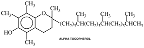
forms: Natural vitamin E contains a variety of tocopherols, including d-Alpha-tocopherol, d-Beta-tocopherol, d-Gamma-tocopherol, d-Delta-tocopherol.
See note below.
tradenames: Trolox®, Aquasol E®
overview of interactions:
• nutrient affecting drug toxicity: Chemotherapy
• nutrients affecting drug toxicity and performance: Antioxidants and Chemotherapy
• nutrient affected by drug: Cholestyramine
• nutrient affected by drug: Clofibrate
• nutrients affected by drug: Colestipol
• Antioxidant nutrients (Vitamin A, Beta-Carotene and Vitamin E) affecting drug performance: Cyclophosphamide
• nutrient affecting drug performance: Cyclosporine
• nutrient affecting drug toxicity: Dapsone
• nutrient affecting drug performance and toxicity: Doxorubicin
(Adriamycin®)
• Alpha-tocopherol affecting drug performance: Griseofulvin
• nutrient affecting drug performance: Haloperidol
• nutrient affected by drug: Lovastatin
• nutrient affecting drug toxicity: Lovastatin
• nutrient affected by drug: Mineral Oil
• nutrient affected by drug: Phenytoin (Dilantin®)
• nutrient affecting drug performance and toxicity: Sulfonylureas
• nutrient affecting drug toxicity: Valproic Acid
• nutrient affecting drug performance: Zidovudine (AZT)
chemistry:
• Tocopherols are oily yellow liquids, water insoluble, heat and acid stable, that deteriorate with exposure to alkali, light, oxygen, and on contact with iron and lead. Frying or foods and freezing also decreases the potency of Vitamin E. However tocopherol esters, which are most commonly found in foods, tend to be fairly resistant to both frying and freezing.
• There are 8 tocopherols. Of these, 4 occur naturally in foods, (alpha, beta, gamma, delta or a, b, g and d ). d-alpha tocopherol accounts for 80% of the activity of the vitamin, however it may exhibit lower in-vivo or in-vitro antioxidant activity when compared to the other less biologically active tocopherols. d,l -alpha tocopherol (SRR a-tocopherol) is the name given to synthetic derivatives which are composed of equal amounts of all the stereoisomers.
• equivalencies:
1 mg d-alpha tocopheryl acetate = 1.49 IU.
synthetic 1 mg d,l-alpha tocopherol = 1 IU.
1 mg d-alpha tocopheryl acetate = 1 alpha tocopherol equivalent
metabolism:
• Vitamin E requires the presence of bile for absorption.
• At normal levels of intake only 20-30% of dietary vitamin E is absorbed. This percentage decreases with increased dosage.
• A diet high in unsaturated fat increases vitamin E requirements.
• Vitamin E is stored in high amounts in the pituitary gland and the adrenals.
• There is no correlation between serum levels and Vitamin E stores. A serum peroxide value can give an indirect status.
• Storage of vitamin E is limited. The liver briefly stores vitamin E but only in small quantities. Adipose tissue and the adrenal glands do store vitamin E as well. The adipose tissue slowly accumulates vitamin E and then in time slowly releases it as well.
function:
• Anti-oxidant: Vitamin E protects cell membranes, especially in the lungs and red blood cells. It particularly protects fatty acids against oxidative damage caused by various pollutants, peroxides, and free radicals formed during metabolic processes. It aids in the prevention of lipofuscin, an oxidized fat that has been implicated in the aging process. If insufficient vitamin E is present, PUFAs may become oxidized in the body. This causes various toxins to be formed. These toxins may lead to chromosomal damage and the formation of cancers. Evidence has been presented that supplemental vitamin E along with vitamin C may greatly inhibit the formation of mutagens in the feces, thus preventing the formation of polyps and cancer. Vitamin E may protect the liver and the rest of the body against environmental pollutants such as ozone and other constituents of smog. People receiving chemotherapy or radiation can also be protected with additional vitamin E supplementation. However, the antioxidant effect of higher dosages of vitamin E may be counterproductive during some forms of chemotherapy and radiation treatment.
• Vitamin E works synergistically with other antioxidant nutrients including selenium, vitamin C, B-carotene and others to quench free radicals, peroxides and other potentially harmful substances. Vitamin E can spare other antioxidants and vice versa.
Red blood cells are particularly susceptible to oxidative damage because of their high oxygen tension. Sickle cell anemia has been successfully treated with vitamin E.
Other antioxidant functions:
• Nerve and muscle function: vitamin E is vital for the protecting of nerve and muscle cell function. In children who had a lack of bile (cholestasis) to absorb fats and vitamin E it was found that vitamin E was able to protect against damage to Schwann cells, dorsal root ganglia and muscle cells. In adults who are deficient in vitamin E peripheral nerve degeneration results.
• Premature infants: develop retrolental fibroplasia or damage to the retina due to their high oxygen exposure. Vitamin E can protect against this damage. In addition vitamin E can protect the lens of the eye from oxidation, cross linking of the collagen fibers and formation of cataracts.
• Muscular dystrophy: type diseases have been treated with vitamin E with only anecdotal success.
• Cholesterol peroxidation: can occur when vitamin E is deficient. In particular LDL cholesterol yields epoxycholesterols which have been implicated in cardiovascular disease.
Other functions of vitamin E:
• Anti-inflammatory effects: vitamin E inhibits the enzyme lipoxygenase, an enzyme responsible for the formation of leukotrienes which cause inflammation. This can be useful in the treatment of asthma and other inflammatory conditions such as arthritis.
• Anti-platelet aggregation effects: vitamin E at higher doses has been shown to increase production of prostaglandin I2 which inhibits platelet stickiness.
dietary sources:
• Unrefined cold-pressed vegetable oils, particularly wheat germ oil and soybean oil, all whole raw or sprouted seeds, nuts, and grains (especially whole wheat), fresh wheat germ (must be absolutely fresh, less than a week old; rancid wheat germ does not contain vitamin E), green leafy vegetables, egg yolks, legumes.
• It is usually difficult to obtain the therapeutic dosage levels typically provided by supplements from food sources alone.
deficiency:
• Vitamin E deficiency is rare in humans. One study, in which mental patients were supplied with 3 mg of vitamin E, resulted in the hemolysis of their red blood cells. It has also been observed that SIDS babies have blood levels of vitamin E similar to those of premature infants. Respiratory stress may be related to lack of antioxidant protection early in life. Premature infants are susceptible to damage of the retina (retrolental fibroplasia) due to the high oxygen tension they are exposed to. Vitamin E can protect against such oxygen generated damage to the retina and also the lungs as well.
• In animal studies central nervous system changes (encephalomalacia) and exudative diathesis occurs. There may also be liver degeneration and reproductive failure affecting both males and females. Muscular dystrophy is another potential sign of a vitamin E deficiency.
• The signs and symptoms of vitamin E deficiency are dry skin, easy bruising, decreased clotting time, eczema, elevated indirect bilirubin, psoriasis, elevated heavy metals, premenstrual syndrome, cystic fibrosis, sickle cell anemia, beta thalassemia, cataracts, fibrocystic disease, benign prostatic hypertrophy, poor wound healing, hot flashes, growing pains, Osgood-Schlatter disease.
• Optimal intake level is dependent upon the intake of unsaturated fatty acids. The greater the content of fatty acids in the diet the greater the requirement of vitamin E. Other antioxidant nutrients can to a certain extent spare vitamin E. The level of exposure to environmental pollutants such as cigarette smoke, industrial pollution and various other chemicals also determines the ideal requirement for vitamin E. People working in areas where their exposure to toxins is greater, such as a bar, toll booth, print shop, nuclear power plant and many others have an increased requirement for vitamin E as well as other antioxidant nutrients. People receiving chemotherapy and/or radiation have an increased requirement because of the damaging effects of these elements to healthy tissue. Intake should be increased before, during and after therapy with these agents. Silymarin has synergistic activity with vitamin E. It is one of the most powerful antioxidants for the liver. It is derived from the plant Silybum marianum.
(Marz R. 183, 1997)
known or potential therapeutic uses: Acne, AIDS/HIV support, allergies, Alzheimer’s disease, anemia (sickle cell anemia and other types of hemolytic anemia), angina, anti-inflammatory, anti-oxidant, atherosclerosis, bronchitis, burns, cardiovascular effects, cataracts, cold sores, cystic fibrosis, diabetes mellitus, Down syndrome, Dupuytren’s contracture, dysmenorrhea, fibrocystic breast disease, fibromyalgia, hepatitis, herpetic lesions and post-herpetic neuralgia, high-altitude exercise performance, high cholesterol, hypoglycemia, immune support, infertility, inflammatory thrombophlebitis, intermittent claudication, macular degeneration, menopause, menorrhagia (heavy menstruation), muscular dystrophy, nocturnal cramping, Osgood-Schlatter disease, osteoarthritis, photosensitivity, premature infants, premenstrual syndrome, psoriasis, Raynaud's syndrome, restless leg syndrome, retinopathy, retrolental fibroplasia, rheumatoid arthritis, scar tissue, seborrheic dermatitis, scleroderma, spontaneous abortion, sudden infant death syndrome (SIDS), sunburn, systemic lupus erythematosis (SLE), tardive dyskinesia, vaginitis, wound healing, yellow nail syndrome.
maintenance dose: 100 - 400 IU per day.
• RDA: 15 IU per day.
• Optimal daily intake: 400 IU.
therapeutic dose: 800 - 3,500 IU per day.
• preferred forms: d-alpha tocopherol and d-alpha tocopheryl (acetate or succinate).
• The tocopherol form may provide slightly higher absorption, while the tocopheryl forms may have slightly longer shelf life. Tocopherol works topically, while tocopheryl does not.
side effects:
• Vitamin E supplements are widely considered to be safe and unlikely to cause adverse side effects in most individuals at typical dosage levels.
• Nausea, flatulence, diarrhea may occur from excessive vitamin E intake.
• Increased triglycerides may result from vitamin E toxicity.
toxicity:
• Vitamin E toxicity is generally considered to be very rare.
• Sudden supplementation in unaccustomed individuals could potentially cause an elevation in blood pressure. Hypertension has been reported to develop in certain people with sensitivities to vitamin E. Wilfrid Shutes has warned that D-a-tocopherol if started at too high a dose can increase the contractility of the heart and of the blood pressure; this may be related to soy allergy. Individuals beginning supplementation with vitamin E may benefit from starting out with low doses, e.g.,100 IU, and gradually increasing the dosage.
• 1800 IU per day has been shown to cause a prolonged blood clotting time. Vitamin E may interfere with vitamin K.
• Reports of infant deaths in 1983 due to parenteral administration of vitamin E were found to be related to the solubilizing agent polysorbate instead of vitamin E. This compound was subsequently banned. In premature infants oral preparation of vitamin E with high osmolality reported cases of necrotizing
enterocolitis.
contraindications:
• Chronic rheumatic heart disease: do not start with high doses, gradually work up.
• Cyclophosphamide (Cytoxan): requires hepatic oxidation for activation; antioxidants interfere with this process.
• Hypertension.
Note: When a label lists mixed tocopherols, it indicates that 80% of the natural d-alpha variety is present with the remaining 20% being a mixture of b, g and d. An important distinction should be made regarding the different forms. The d alpha tocopheryl has
the most biological activity of all the forms (currently this stereoisomer is now referred to as RRR-a-tocopherol where the Rs refer to the stereoisomers of tocopherol). This means that it will prevent deficiency symptoms in laboratory animals chicks and rats more efficiently than any of the other form. However, there is a question as to whether animal assays are appropriate for evaluation of human requirements. In addition merely preventing deficiency symptoms may not be the best way to assess the function of tocopherol or for that matter other nutrients as well. Where one tocopherol form may prevent deficiency symptoms, another, with less biological activity may be better at preventing oxidation of LDL cholesterol or lipid membranes and at lowering serum cholesterol, which is the case of g tocopherol. The tocotrienols, which are less distributed in nature than the tocopherols, have not been well studied probably because they have so-called less biological activity. However one study reported that it has greater antitumor effects than the most biological active tocopherol.
(Komiyama K, et al. Chem Pharm Bull 1989;37, 1369-1381.)
Interactions
nutrients affecting drug toxicity: Vitamin A and Vitamin E for Mouth Sores due to Chemotherapy
• mechanism: Chemotherapeutic agents are responsible for a wide variety of adverse effects in patients receiving them due to damage of healthy cells and derangement of normal metabolic processes. Vitamin A and vitamin E are well known for their abilities to enhance restoration of healthy cells and tissues.
• adverse drug effects: Mouth sores are a common side effect of chemotherapy treatment.
• research: Mills examined the beneficial effect provided by beta-carotene on mouth sores, specifically oral mucositis, induced by radiation and chemotherapy induced oral mucositis. When compared to controls receiving equivalent radiation and chemotherapy, subjects receiving alpha-tocopherol, 400,000 IU daily for three weeks followed by 125,000 IU for four weeks, experienced slower onset and decreased severity of mouth inflammation. In a later randomized, double-blind, placebo-controlled study involving 18 patients Wadleigh et al used topical vitamin E in the treatment of oral mucositis in patients receiving chemotherapy. They found that six of nine patients receiving vitamin E had complete resolution of their oral lesions while lesions did not resolve completely in eight of nine patients who received placebo. Lopez et al conducted further studies looking at the efficacy of topical vitamin E for the treatment of this condition and found that while topical vitamin E, applied once daily, was beneficial to some subjects, others did not experience relief from their symptoms.
(Mills EED. Brit J Cancer 1988 Apr;57(4):416-417; Wadleigh RG, et al. Am J Med
1992 May;92(5):481-484; Lopez I, et al. Ann Med Interne (Paris) 1994;145(6):405-408.)
• nutritional support: While the research thus far has not been conclusive many individuals suffering from mouth sores due to chemotherapy would probably experience decreased symptoms and faster healing through the application of vitamin E. Such treatment carries no significant risks and has been shown to help some patients. In such cases topical application of alpha tocopherol twice daily would be the form most likely to be effective; with alpha tocopheryl being the less preferred form.
nutrient affecting drug toxicity: Alpha-tocopherol and cardiac toxicity due to Chemotherapy
• research: Many chemotherapeutic agents, most notably adriamycin, are used in less than optimal doses due to their toxic effects upon the heart. In 1979 Krivit proposed that alpha-tocopherol might ameliorate or prevent cardiac dysfunction without impairing the drug's antitumor effectiveness. Subsequently a study by Legha et al showed that use of the vitamin did not compromise the drug's antitumor activity but also failed to demonstrate substantial protection against adriamycin-induced cardiac toxicity.
(Krivit W. Am J Pediatr Hematol Oncol 1979 Summer;1(2):151-153; Legha SS, et al.
Ann NY Acad Sci 1982;393:411-418.)
• nutritional support: Until more positive research findings emerge no substantial evidence exists to support the use of alpha-tocopherol as a method of reducing cardiac toxicity due to chemotherapeutic agents such as
adriamycin.
nutrient affecting drug toxicity and performance: Antioxidants
and Chemotherapy
• mechanism: Oxygen radicals have increasingly come under investigation as highly toxic stressors contributing to the causes of many disease, including many forms of cancer. Vitamin A and Vitamin C are well known antioxidant agents. N-acetylcysteine (NAC) is a free radical scavenger and might access the endothelial cell thus increasing intracellular glutathione (GSH) stores. Even so, some concerns have been raised antioxidants might be contraindicated during chemotherapy because one way in which chemotherapeutic agents attack cancer cells is by causing oxidative damage.
(Weijl NI, et al. Ann Oncol 1998 Dec;9(12):1331-1337; Kong Q, Lillehei KO.
Med Hypotheses 1998 Nov;51(5):405-409.)
• research: A variety of test tube-based and animal studies have found vitamin A, C and E to increase the effectiveness of chemotherapy in several kinds of cancer. NAC therapy may be useful therapy in advanced cervical cancers, especially squamous cell carcinomas. In a double-blind study Wagdi et al found that an antioxidant combination consisting of vitamin C, vitamin E, and N-acetylcysteine (NAC) provided protected against heart damage induced by chemotherapy without reducing with the drug's effectiveness. Several researchers have offered the seemingly paradoxical conclusion that the appropriate administration of antioxidant inhibitors and/or free-radical-generating compounds may be a useful strategy in the treatment of solid tumors. After performing a comprehensive review of the relationship between chemotherapy and antioxidants Weijl et al rebuffed warnings that antioxidants needed to be avoided during chemotherapy but also determined that there substantive evidence was lacking to support the use of antioxidants to provide relief from the adverse side effects of chemotherapy. More recently however, conference reports by Salignik R, and co-worker Zeisel SH, at a recent American Society of Cell Biology meeting suggest that Vitamin A and Vitamin E deficient mice were less susceptible to brain tumor progression than non vitamin-deficient control animals. (unpublished proceedings) Salignik suggested suppression of free radicals by the anti-oxidant vitamins may suppress apoptosis (programmed cell death).
(Sacks PG, et al. Int J Cancer 1995;61:409-415; Taper HS, et al. Int J Cancer 1987;40:575-579; Kurbacher CM, et al.
Cancer Letters 1996:103-119; Gillissen A, Nowak D. Respir Med 1998 Apr;92(4):609-623; Fujiwaki R, et al.
Anticancer Res 1997 Sep-Oct;17(5B):3751-3755.Wagdi P, et al. Jpn Heart J 1996;37:353-359; Weijl NI, et al.
Cancer Treatment Rev 1997;23:209-240; Kong Q, Lillehei KO. Med Hypotheses 1998 Nov;51(5):405-409; Domenighetti G, et al.
Rev Mal Respir 1999 Feb;16(1):29-37; Kodama J, et al. Gan To Kagaku Ryoho 1999 Jan;26(1):89-92; Albini A, et al.
Int J Cancer 1995 Mar 29;61(1):121-129; Weijl NI, et al. Cancer Treatment Rev 1997 Jul;23(4):209-240; Salignik R, et al.
39th Annual Meeting of the American Society for Cell Biology, Dec 11-15th, 1999.)
• nutritional concerns: Concerns have been raised by some oncologists that supplementation with antioxidants might in some way interfere with or limit the effectiveness of chemotherapeutic agents. However, no substantial research has emerged to support this speculation or to warrant considering antioxidants as contraindicated during the course of chemotherapy. Many nutritionally oriented healthcare professionals consider the oxidative damage caused by chemotherapy to be a particularly troublesome side effect given that evidence increasingly points to oxidative damage as being a contributing factor in the causation of many cancers. Individuals receiving chemotherapy should consult their treating physician and/or a nutritionally trained healthcare professional about potential value of adding antioxidants to their regime before starting such supplementation.
nutrient affected by drug: Cholestyramine
• mechanism: Bile acid sequestrants, such as cholestyramine, decrease lipid digestion and absorption, as well as absorption of the fat-soluble vitamins and other nutrients.
(Roe DA. 1985: 158-159; Watkins DW, et al. Dig Dis Sci 1985 May;30(5):477-482; Hathcock
JN. Fed Proc. 1985 Jan;44(1 Pt 1):124-129; West RJ, Lloyd JK. Gut. 1975 Feb;16(2):93-98.)
• nutritional support: Regular use of a high-potency multiple vitamin/mineral will replace the nutrients impeded by the drug.
nutrient affected by drug: Clofibrate
• mechanism: Clofibrate impairs absorption of vitamin E.
(Vitamin E Fact Book. 1994.)
nutrient affected by drug: Colestipol; along with Vitamin A, Vitamin D, Vitamin K and Folate
• mechanism: Colestipol, by design, reduces fat absorption and hence interferes with absorption of fat-soluble nutrients.
(Tonstad S, et al. Arch Dis Child 1996 Feb;74(2):157-160.)
• clinical concerns: This interaction raises serious questions given the accumulating experimental, epidemiological, and clinical evidence of an association between anti-oxidant vitamin intake (especially vitamin E) and reduced risk of coronary heart disease.
• research: Hodis et al have reported an association between supplementary vitamin E intake and angiographically demonstrated reduction in coronary artery lesion progression. Tonstad et al conducted a study of 37 boys and 29 girls aged 10-16 years with familial hypercholesterolaemia first in an eight week double blind, placebo controlled protocol, then in open treatment for 44-52 weeks. They found that levels of serum folate, vitamin E, and carotenoids were reduced in the colestipol group. After one year of colestipol, those who took 80% or more of the prescribed dose had a greater decrease in serum 25-hydroxyvitamin D levels than those who took less than 80%.
(Hodis HN, et al. JAMA 1995 Jun 21;273(23):1849-1854; Tonstad S, et al. Arch Dis Child 1996 Feb;74(2):157-160.)
• related research: While supplementary vitamin E has been found to be effective in treating high cholesterol by reducing progression of carotid arterial wall intima-media thickness, there has been no confirmation that the addition of moderate doses of vitamin E along with colestipol will enhance the effectiveness of the therapy in reducing the progression of atherosclerosis.
(Azen SP, et al. Circulation 1996 Nov 15;94(10):2369-2372; Schlierf G, et al.
Klin Wochenschr. 1985 Sep 2;63(17):802-8026; Schwarz KB, et al. Pediatrics. 1980 Feb;65(2):243-250; Leonard JP, et al.
Arzneimittelforschung 1979;29(7):979-981; Tsang RC, et al. Pediatr Res. 1978 Oct;12(10):980-982.)
• nutritional support: Vitamin E supplementation, typically 400-800 IU per day, would most likely benefit individuals taking
colestipol.
Antioxidant nutrients (Vitamin A, Beta-Carotene and Vitamin E) affecting drug performance: Cyclophosphamide
• research: Preliminary studies on humans undergoing chemotherapy with cyclophosphamide have indicated promise of increased survival rates when supplemented with antioxidant nutrients. While antioxidants might have many benefits as supplements, evidence as to their effectiveness in patients taking cyclophosphamide are inconclusive.
(Jaakkola K, et al. Anticancer Res 1992 May;12(3):599-606; Venugopal M, et al.
Life Sci 1996;59(17):1389-1400; Vinitha R, et al. Jpn J Med Sci Biol 1995 Jun;48(3):145-156.)
• nutritional concerns: Since activation of cyclophosphamide requires oxidation in the liver there has been concern that antioxidant supplementation might interfere with its effectiveness. However, at this time such concern remains speculative as no conclusive evidence as emerged to confirm an adverse interaction of this type.
(Labriola D, Livingston R. Oncology (Huntingt). 1999 Jul;13(7):1003-1008.)
nutrient affecting drug performance: Cyclosporine
• mechanism: Several studies have found that 25 mg/kg of "water-soluble" d-a vitamin E increases the absorption of cyclosporine and may decrease the needed dose of cyclosporine by 40-72% without reducing its effectiveness.
(Chang T, et al. Clin Pharmacol Ther 1996 Mar;59(3):297-303; Sokol RI et al.
Lancet 1991;338:212-215; Pan SH, et al. Pharmacotherapy 1996 Jan;16(1):59-65.)
• nutritional synergy: Supplementing 25 IU of vitamin E per 2.2 pounds of body weight is an appropriate dosage as indicated by the research. However, since supplementation with vitamin E will effect the drug's potency and dosage, any such usage should be done in consultation with the prescribing doctor, and preferably in collaboration with a nutritionally-oriented physician. Apart from some potential for reduced toxicity, the most obvious value in this increased absorption derives from the reduced cost of the therapy as cyclosporine is very expensive.
nutrient affecting drug toxicity: Dapsone
• mechanism: Dapsone is strongly oxidative in a way that damages the membranes of red cells and results in hemolysis.
• research: In several trials, individuals, with leprosy or dermatitis herpetiformis, taking Dapsone were given 800 IU of vitamin E for periods ranging from four weeks to three months. In one study of patients with dermatitis herpetiformis, vitamin E therapy was followed by vitamin C therapy, and then combined vitamins E and C. This study concluded that the vitamin C had exerted no beneficial effect but that oral administration of 800 units of vitamin E daily for 4 weeks confers partial protective effect against dapsone-induced hemolysis in patients with dermatitis herpetiformis.
(Kelly JW, et al. Arch Dermatol 1984 Dec;120(12):1582-1584; Prussick R, et al.
Arch Dermatol 1992 Feb;128(2):210-213; Lardo MM, et al. Medicina (B Aires) 1997;57(2):150-154.)
• nutritional support: The clinical significance of vitamin E to counter the adverse effects of Dapsone remains somewhat inconclusive. However, supplementation of 800 IU would provide many other potential benefits for such individuals beyond the anti-oxidant effect indicated in these studies.
nutrient affecting drug performance and toxicity: Doxorubicin
(Adriamycin®)
• mechanism: Antioxidant action reduces cardiac toxicity.
• research: Initial research by Prasad et al suggested that supplementation with vitamin E might reduce cardiac and skin toxicity due to doxorubicin. Research studies with animals have found that vitamin E's potent antioxidant activity can protect against Adriamycin-induced cardiotoxicity; hence reducing the risk of heart failure which is a serious side effect associated with doxorubicin.
(Prasad KN, et al. Proc Soc Exp Biol Med 1980 Jun;164(2):158-163; Ellison NM.
Cancer Bull 1985;37(3):112-113; Am Heart J 1986;lll:95; Myers C, et al. Cancer Treat Rep 1976;60:961-962; Sonneveld P.
Cancer 1978;62:1033-1036.)
However, no conclusive evidence has come forth confirming the cardioprotective effect of vitamin E in human trials. Nevertheless, some evidence suggests that supplementation with vitamin E may allow use of higher doses of doxorubicin without correspondingly increasing toxicity.
(Ellison, NM, Londer H. 1981; Weiji NI, et al. Cancer Treatment Rev 1997,23:209-210.)
Anecdotal reports indicate that very high doses of vitamin E (1600 IU) may reduce the amount of alopecia (hair loss) resulting from use of doxorubicin. (Wood LA.
N Engl J Med 1985;312:1060 ) As of yet, no research on humans has duplicated the protective effect against hair loss found in one study with rabbits.
(Weiji NI, et al. Cancer Treatment Rev 1997;23:209-240.)
• nutritional support: Supplementation of vitamin E at doses of 800 IU or more is prescribed by some practitioners as nutritional support for patients taking doxorubicin, even though no decisive evidence has emerged showing that the vitamin reduces drug toxicity or protects against hair loss.
• nutritional synergy: In vitro evidence suggests that vitamin E enhances the growth inhibitory effect of doxorubicin, at least in a test tube.
(Ripoll EAP, et al. J Urol 1986;136:529-531.)
Alpha-tocopherol affecting drug performance: Griseofulvin
• mechanism: Reduction of P-450 system enzyme activities in the liver achieved with alpha-tocopherol slows down the biotransformation rate of griseofulvin and significantly elevates its blood serum and skin concentrations.
(Lashmanova AP, et al. Vestn Dermatol Venerol 1990;9:15-16.)
• nutritional synergy: Researchers found that by supplementing 50 IU of vitamin E per day in children on griseofulvin blood levels of the drug within four weeks increased enough to allow the drug dose to be reduced by half. While this reduced dose would be beneficial in reducing the drug's side effects, patients prescribed griseofulvin should not supplement with vitamin E or reduce their dosage of the drug without consulting their prescribing physician.
(Mycol Observer. Nov/Dec 1990:8; Leshchenko VM, et al. Vestn Dermatol
Venerol. 1988;(12):24-26; Omel'chenko OG. Vestn Dermatol Venerol. 1987;(8):12-15.)
nutrient affecting drug performance: Haloperidol
• mechanism: Haloperidol is cytotoxic to numerous parts of the brain, specifically the primary hippocampal neurones, C6 glioma cells and NCB20 cells. Researchers have investigated the possibility that various activities of vitamin E, particularly as a free radical scavenger, might reduce the toxic side effects associated with use of the drug.
• research: In vitro research indicates that chronic haloperidol administration can reduce striatal vitamin E levels. In other test tube studies looking at haloperidol-induced toxicity Behl et al determined that vitamin E (alpha-tocopherol), a lipophilic free radical scavenger, prevented DNA fragmentation and ultimately cell death. Based upon their research involving rats Gattaz et al suggested that the concomitant administration of vitamin E to neuroleptics might prevent the development of tardive dyskinesia in humans. However, in human studies supplementation with vitamin E neither provides protective effect against haloperidol-induced symptoms nor does it interfere with the therapeutic efficacy of the drug.
(VonVoigtlander PF, et al. Res Commun Chem Pathol Pharmacol. 1990 Jun;68(3):343-352; Gattaz WF, et al.
J Neural Transm Gen Sect 1993;92(2-3):197-201; Behl C, et al. Neuroreport 1995 Dec 29;7(1):360-364; Eranti VS, et al.
Psychopharmacology (Berl). 1998 Dec;140(4):418-420.)
nutrient affected by drug: Lovastatin
• metabolic/nutrient concern: Alpha-tocopherol is a potent endogenous antioxidant. Much attention has been given to its role in reducing the risk of atherosclerosis based on the theory that the pathological changes result from oxidative processes. Likewise alpha-tocopherol is often used in the treatment of cardiovascular disease.
• research: Chen et al conducted initial research into the antiatherosclerotic effects of vitamin E, through preservation of endogenous antioxidant activity and inhibition of lipid peroxidation, when used with lovastatin and amlodipine. Later, in a randomized, double-masked, crossover clinical trial Palomaki et al evaluated whether lovastatin therapy (60 mg daily) affected the initiation of oxidation of low density lipoprotein (LDL) in cardiac patients on alpha-tocopherol supplementation therapy. Twenty-eight men with verified coronary heart disease and hypercholesterolemia received 450 IU alpha-tocopherol daily with lovastatin or with placebo. They concluded that alpha-tocopherol supplementation significantly increased the antioxidative capacity of LDL when measured ex vivo, but that this benefit was partially abolished by concomitant lovastatin therapy.
(Chen L, et al. J Am Coll Cardiol. 1997 Aug;30(2):569-575; Palomaki A, et al.
Arterioscler Thromb Vasc Biol 1999 Jun;19(6):1541-1548.)
• nutritional support: 400 IU of alpha-tocopherol, twice daily, would be an appropriate dose to counter the adverse effects of lovastatin and provide some additional benefit to the cardiovascular system. Individuals who are also taking lovastatin should consult with their prescribing physician and/or a nutritionally oriented healthcare professional to supervise and monitor the course of treatment.
nutrient affecting drug toxicity: Vitamin E
• mechanism: As discussed above lovastatin has been found to deplete Coenzyme Q10.
• nutritional support: Vitamin E supplementation of may offset the loss of Coenzyme Q10. 400 IU of alpha-tocopherol, twice daily, would be an appropriate dose.
(Baum H. New Scientist May 24, 1991, 24.)
nutrient affected by drug: Mineral Oil
• mechanism: Mineral oil, as a lipid solvent, may also absorb many substances and/or interfere with normal absorption of alpha-tocopherol and other nutrients.
• research: While there is some disagreement, most research has found that mineral oil interferes with the absorption of many nutrients, including beta-carotene, calcium, phosphorus, potassium, and vitamins A, D, K, and E. Mineral oil, as a lipid solvent, may also absorb many substances. Chronic use of mineral oil can cause a deficiency of vitamins A, D, E, and K, being fat soluble, as a result of their being absorbed.
(Clark JH, et al. Am J Dis Child 1987 Nov;141(11):1210-1212; Holt GA.1998, 176.)
• nutritional concerns: If using mineral oil for any extended period of time, regular use of a multivitamin supplement would be beneficial. Malabsorption of fat-soluble vitamins due to ingestion of mineral oil can be minimized by administering mineral oil on an empty stomach or consuming vitamin or mineral supplements at least two hours before or after the mineral oil. In general it is advisable to limit the internal use of mineral oil to periods of less than 1 week.
nutrient affected by drug: Phenytoin (Dilantin®)
• research: Phenytoin reduces serum levels of vitamin E.
(Sullivan, C, et al. Med J Austr 1990 Jun 4;152(11):613-614.)
• nutritional support: Supplementing with vitamin E has been reported by some researchers to reduce the incidence of seizures, most likely due to its antioxidant activities. However, others have not been able to confirm these findings of independent anticonvulsant effects from vitamin E. Other clinical studies have found vitamin E (d-alpha-tocopherol) to be a useful adjunct to anticonvulsant drugs. Improvement has been reported even in patients with complex partial seizures, which are often resistant to drug therapy.
(Wiehl W, Hart LL. DICP 1991 Apr;25(4):362-363; Sullivan, C, et al. Med J Austr 1990 Jun 4;152(11):613-614; Raju GB, et al.
Epilepsia 1994 Mar-Apr;35(2):368-732.)
nutrient affecting drug performance and toxicity: Sulfonylureas
• mechanism: Vitamin E supplements suppress hemoglobin glycation. Consequently, glycemic control may appear falsely exaggerated.
(Drug Evaluations Subscription. Winter, 1994.)
nutrient affecting drug toxicity: Valproic Acid
• mechanism: Specific oxidative metabolites of valproic acid have been associated with the drug's toxicity.
• research: Research indicates that the valproic acid's cytotoxic activity is the result of generation of hydrogen peroxide and the production of highly reactive hydroxyl free radicals. Graf et al looked at children with serious adverse experiences with valproic acid and found that glutathione peroxidase was significantly depressed and glutathione reductase was significantly elevated relative to other subjects. In particular, they reported that selenium and zinc concentrations were lower in a serious adverse experience patients than in controls and concluded that selenium dependent antioxidant activity might play a special role protecting against adverse reactions. Buchi et al found that the free-radical scavenging action of alpha-tocopherol (vitamin E) and N,N'-diphenyl-p-phenylenediamine (DPPD) protected against lipid peroxidation and hepatotoxicity caused by valproic acid (VPA) in rats.
(Nurge ME, et al. Nutr Res 1991;11:949-960; Tabatabaei AR, et al. Toxicology 1996 Aug 1;112(1):69-85; Abbott FS.
Chem Res Toxicol 1999 Apr;12(4):323-330; Graf WD, et al. Neuropediatrics 1998 Aug;29(4):195-201; Buchi KN, et al.
J Clin Pharmacol 1984 Apr;24(4):148-154.)
• nutritional support: While the use of valproic acid produces numerous adverse effects, especially upon the liver, no conclusive evidence has emerged to demonstrate the clinical role of vitamin E and selenium, or other antioxidants, in countering these adverse effects. Individuals taking valproic acid should consult their prescribing physician and/or a nutritionally trained healthcare professional regarding the use of antioxidants as part of a nutritional support program.
nutrient affecting drug performance: Zidovudine (AZT)
• research: Test tube studies suggest that supplementation with vitamin E (alpha-tocopherol) may enhance the therapeutic efficacy of AZT.
(Gogu SR, et al. Biochem Biophys Res Commun. 1989 Nov 30;165(1):401-407; Gogu SR, et al.
Exp Hematol. 1991 Aug;19(7):649-652; Odeleye OE, Watson RR. Prog Food Nutr
Sci. 1991:15(1-2):1-19.)
• nutritional support: Supplementation of vitamin E at doses of 800 IU might provide nutritional support for patients taking zidovudine, even though no decisive evidence has emerged showing that the vitamin reduces drug toxicity or enhances its efficacy. Individuals on a regime including zidovudine should consult their prescribing physician and/or a healthcare professional trained in nutritional therapy before starting megadoses of any supplements.


Please read the disclaimer concerning the intent
and limitations of the information provided here.
Do not rely solely on the information in this article.
The information presented in Interactions is for
informational and educational purposes only. It is based on scientific
studies (human, animal, or in vitro), clinical experience, case
reports, and/or traditional usage with sources as cited in each
topic. The results reported may not necessarily occur in all
individuals and different individuals with the same medical conditions
with the same symptoms will often require differing treatments. For
many of the conditions discussed, treatment with conventional medical
therapies, including prescription drugs or over-the-counter
medications, is also available. Consult your physician, an
appropriately trained healthcare practitioner, and/or pharmacist for
any health concern or medical problem before using any herbal products
or nutritional supplements or before making any changes in prescribed
medications and/or before attempting to independently treat a medical
condition using supplements, herbs, remedies, or other forms of
self-care.
References
[No author given.] Drug Evaluations Subscription. Chicago: American Medical Association, Vol. II, Section 10, Chapter 3, Winter, 1994.
[No authors listed] MRC/BHF Heart Protection Study of cholesterol-lowering therapy and of antioxidant vitamin supplementation in a wide range of patients at increased risk of coronary heart disease death: early safety and efficacy experience.
Eur Heart J. 1999 May;20(10):725-741.
Abstract: AIMS: In observational studies, prolonged lower blood total cholesterol levels - down at least to 3 mmol. l-1 - are associated with lower risks of coronary heart disease. Cholesterol-lowering therapy may, therefore, be worthwhile for individuals at high risk of coronary heart disease events irrespective of their presenting cholesterol levels. Observational studies also suggest that increased dietary intake of antioxidant vitamins may be associated with lower risks of coronary heart disease. The present randomized trial aims to assess reliably the effects on mortality and major morbidity of cholesterol-lowering therapy and of antioxidant vitamin supplementation in a wide range of different categories of high-risk patients. METHODS AND RESULTS: Men and women aged 40 to 80 years were eligible provided they were considered to be at elevated risk of coronary heart disease death because of past history of myocardial infarction or other coronary heart disease, occlusive disease of non-coronary arteries, diabetes mellitus or treated hypertension; had baseline blood total cholesterol of 3.5 mmol. l-1 or greater; and no clear indications for, or contraindications to, either of the study treatments. Eligible patients who completed a pre-randomization run-in phase on active treatment were randomly allocated to receive simvastatin (40 mg daily) or matching placebo tablets and, in a '2x2 factorial' design, antioxidant vitamins (600 mg vitamin E, 250 mg vitamin C and 20 mg beta-carotene daily) or matching placebo capsules. Follow-up visits after randomization are scheduled at 4, 8 and 12 months, and then 6-monthly, for at least 5 years.Between July 1994 and May 1997, 15 454 men and 5082 women were randomized, with 9515 aged over 65 years at entry. Diagnostic criteria overlapped, with 8510 (41%) having had myocardial infarction (most of whom were either female, or elderly or with low blood cholesterol), 4869 (24%) some other history of coronary heart disease, 3288 (16%) cerebrovascular disease, 6748 (33%) peripheral vascular disease, 5963 (29%) diabetes mellitus (of whom 3985 had no history of coronary heart disease) and 8455 (41%) treated hypertension. Baseline non-fasting total cholesterol levels were less than 5.5 mmol. l-1 in 7882 (38%) participants, and LDL (low density lipoprotein) cholesterol less than 3.0 mmol. l-1 in 6888 (34%).During a mean follow-up of 25 months (range: 13 to 47 months), no significant differences had been observed between the treatment groups in the numbers of patients with muscle symptoms, other possible side-effects leading to termination of study treatment, or elevated liver and muscle enzymes. After 30 months of follow-up, 81% of randomized patients remained compliant with taking their study simvastatin or placebo tablets, and allocation to simvastatin produced average reductions in non-fasting blood total and LDL cholesterol of about 1.5-1.6 mmol. l-1 and 1.1-1.2 mmol. l-1 respectively. Eighty-seven per cent of patients remained compliant with taking their vitamin or placebo capsules, and allocation to the vitamin supplement produced an average increase in plasma vitamin E levels of about 24 micromol. l-1. Based on this initial follow-up period, the estimated annual rate of non-fatal myocardial infarction or fatal coronary heart disease is 2.4%, annual stroke rate is 1.3%, and annual all-cause mortality rate is 2. 2%. CONCLUSION: The Heart Protection Study is large, it has included a wide range of patients at high risk of vascular events, and the treatment regimens being studied are well-tolerated and produce substantial effects on blood lipid and vitamin levels. The study should, therefore, provide reliable evidence about the effects of cholesterol-lowering therapy and of antioxidant vitamin supplements on all-cause or cause-specific mortality and major morbidity in a range of different categories of individuals for whom uncertainty remains about the balance of benefits and risks of these treatments.
Anonymous. Vitamin E and cell injury. Nutr Rev 1988;46:1367. (Review)
Anonymous. Vitamin E boosts griseofulvin. Mycol Observer. Nov/Dec 1990:8. (Letter)
Ayres S Jr, Mihan R. Nocturnal leg cramps (systremma): a progress report on response to vitamin E.
South Med J. 1974 Nov;67(11):1308-1312.
Abstract: 103/125 patients with nocturnal leg and foot cramps, over half of whom had suffered for over 5 years, reported complete or nearly complete relief. 20/125 had a moderate to good response and only 2 failed to respond. l/2 responded to 300 I.U. or less, while half required 400 I.U. or more. Many had to continue vitamin E to prevent cramps from recurring. Response was usually within 1 week. Similar success was obtained with nocturnal rectal cramps, abdominal muscle cramps, and cramps following heavy exercise.
Azen SP, Qian D, Mack WJ, Sevanian A, Selzer RH, Liu CR, Liu CH, Hodis HN. Effect of supplementary antioxidant vitamin intake on carotid arterial wall intima-media thickness in a controlled clinical trial of cholesterol lowering.
Circulation 1996 Nov 15;94(10):2369-2372.
Abstract: BACKGROUND: There is accumulating experimental, epidemiological, and clinical evidence of an association between anti-oxidant vitamin intake and reduced risk of coronary heart disease. Using data from the Cholesterol Lowering Atherosclerosis Study (CLAS), we explored the association of self-selected supplementary antioxidant vitamin intake on the rate of progression of early preintrusive atherosclerosis. METHODS AND RESULTS: CLAS was an arterial imaging trial in which nonsmoking 40- to 59-year-old men with previous coronary artery bypass graft surgery were randomized to colestipol/niacin plus diet or placebo plus diet. The rate of progression of early preintrusive atherosclerosis was determined in 146 subjects using high-resolution B-mode ultrasound quantification of the distal common carotid artery far wall intima-media thickness (IMT). From the nutritional supplement database, 22 subjects had an on-trial average supplementary vitamin E intake of > or = 100 IU per day (high users) and 29 subjects had an average on-trial supplementary vitamin C intake of > or = 250 mg per day (high users). Within the placebo group, less carotid IMT progression was found for high supplementary vitamin E users when compared with low vitamin E users (0.008 versus 0.023 mm/y, P = .03). No effect of vitamin E within the drug group was found. No effect of vitamin C within the drug or placebo group was found. CONCLUSIONS: Supplementary vitamin E intake appears to be effective in reducing the progression of atherosclerosis in subjects not treated with lipid-lowering drugs while the process is still confined to the arterial wall (early preintrusive atherosclerosis).
Baum H. New Scientist May 24, 1991, 24.
Baum M, Cassetti L, Bonvehi P, Shor-Posner G, Lu Y, Sauberlich H. Inadequate dietary intake and altered nutrition status in early HIV-1 infection.
Nutrition 1994 Jan-Feb;10(1):16-20.
Abstract: Recent studies indicate that multiple nutritional abnormalities occur relatively early in the course of human immunodeficiency virus (HIV-1) infection. Decreased plasma levels of vitamins B6, B12, A, and E and zinc have been correlated with dietary intake and associated with significant alterations in immune response and cognitive function. To determine the level of intake consistent with normal plasma nutrient levels, we examined nutrition status in relation to food consumption and nutrient supplementation in HIV-1-seropositive (HIV+) and -seronegative (HIV-) homosexual men. The mean level of total intake (diet plus supplements) for all nutrients was significantly higher in HIV+ men. To achieve normal plasma nutrient values, the HIV+ men appeared to require intake in multiples of the recommended dietary allowance (RDA) for vitamins A, E, B6, and B12 and zinc. For the HIV+ men, a relatively high proportion of biochemical deficiency was associated with consumption of vitamin B6 and zinc at the RDA level. Because little evidence of deficiency was observed with elevated intake in both groups, an effective program of nutritional supplementation may be beneficial in maintaining adequate plasma nutrient levels.
Bays HE, Dujovne CA. Drug interactions of lipid-altering drugs. Drug Saf 1998 Nov;19(5):355-371. (Review)
Behl C, Rupprecht R, Skutella T, Holsboer F. Haloperidol-induced cell death--mechanism and protection with vitamin E in vitro.
Neuroreport 1995 Dec 29;7(1):360-364.
Abstract: Haloperidol, a dopamine receptor antagonist and sigma-receptor-active neuroleptic drug, is cytotoxic to primary hippocampal neurones, C6 glioma cells and NCB20 cells. A 24 h challenge of these cells with haloperidol resulted in reduced cell viability and ultimately cell lysis. The most dramatic changes in cellular morphology were the retraction of cellular extensions, development of membrane blebs, and finally cell detachment from the culture dish. DNA isolated from haloperidol-treated cells was randomly degraded, indicating a necrotic rather than an apoptotic pathway of cell death. Vitamin E (alpha-tocopherol), a lipophilic free radical scavenger, prevented haloperidol-induced DNA fragmentation and ultimately cell death. These findings suggest that haloperidol induces necrotic cell death in which free radicals play a major role.
Beyer RE. The role of ascorbate in antioxidant protection of biomembranes: interaction with vitamin E and coenzyme Q.
J Bioenerg Biomembr. 1994 Aug;26(4):349-358. (Review)
Abstract: One of the vital roles of ascorbic acid (vitamin C) is to act as an antioxidant to protect cellular components from free radical damage. Ascorbic acid has been shown to scavenge free radicals directly in the aqueous phases of cells and the circulatory system. Ascorbic acid has also been proven to protect membrane and other hydrophobic compartments from such damage by regenerating the antioxidant form of vitamin E. In addition, reduced coenzyme Q, also a resident of hydrophobic compartments, interacts with vitamin E to regenerate its antioxidant form. The mechanism of vitamin C antioxidant function, the myriad of pathologies resulting from its clinical deficiency, and the many health benefits it provides, are reviewed.
Buchi KN, Gray PD, Rollins DE, Tolman KG. Protection against sodium valproate injury in isolated hepatocytes by alpha-tocopherol and
N,N'-diphenyl-p-phenylenediamine. J Clin Pharmacol 1984 Apr;24(4):148-154.
The possibility that lipid peroxidation is involved in valproic acid (VPA) hepatotoxicity was explored by testing the ability of the free-radical scavengers alpha-tocopherol (vitamin E) and N,N'-diphenyl-p-phenylenediamine (DPPD) to protect against VPA toxicity. Rat hepatocyte cultures were treated with toxic doses of VPA, in conjunction with varying doses of vitamin E and DPPD. Lactate dehydrogenase (LDH) release into the culture media was used to calculate an LDH index as a measure of toxicity. Vitamin E afforded increasing protection against VPA toxicity at concentrations of 1.0 to 4.0 microM but then leveled off and did not give complete protection at concentrations up to 8.0 microM. No protection was seen at less than 1.0 microM. DPPD showed increasing protection from 0.05 to 0.50 microM, with complete protection at the highest concentration. These data indicate that VPA toxicity can be prevented by simultaneous administration of free-radical scavengers and support the concept that VPA hepatotoxicity is due to lipid peroxidation.
Butler, McKnight. Vitamin E in the primary treatment of primary
dysmenorrhea. Lancet 1955; 1:844-847.
Abstract: 100 women with spasmodic dysmenorrhea received either alpha tocopherol tabs 50mg t.i.d. or placebo for 10 days premenstrually and for the next 4 days. After 2 cycles, 34/50 (68%) in the experimental group improved compared to 9/50 (18%) of the controls.
Chang T, Benet LZ, Hebert MF. The effect of water-soluble vitamin E on cyclosporine pharmacokinetics in healthy volunteers.
Clin Pharmacol Ther 1996 Mar;59(3):297-303.
Abstract: We evaluated the effect of water-soluble vitamin E (d-alpha-tocopheryl polyethylene glycol 1000 succinate [TPGS]; Liqui-E) on the oral pharmacokinetics of the cyclosporine, a poorly available (approximately 30%) drug, in healthy volunteers. Ten healthy subjects were given two doses of oral cyclosporine (10mg/kg) separated by a 7-day washout period. Oral TPGS (2.6 IU/kg) was administered concomitantly with one of the cyclosporine doses in a randomized order. A significant increase was observed in area under the blood concentration-time curve (AUC;mean +/ SD) with concomitant TPGS administration (3908 +/- 2601 versus 6296 +/- 5102 ng x hr/ml). Significant decreases were observed in apparent oral clearance (0.24 +/- 0.14 versus 0.15 +/- 0.08 L/hr/kg) and apparent oral steady-state volume of distribution (1.57 +/- 0.95 versus 1.07 +/- 0.73 L/kg). No significant changes were observed in the ratios of metabolites to parent drug AUC values. The comparable relative decreases in apparent oral clearance (38%) and apparent oral steady-state volume of distribution (30%) with TPGS are most likely explained by enhanced absorption, decreased counter transport back into the intestine by P-glycoprotein, or some unknown mechanism by which cyclosporine is protected from metabolism in the gut, thereby increasing bioavailability.
Chen L, Haught WH, Yang B, Saldeen TG, Parathasarathy S, Mehta JL. Preservation of endogenous antioxidant activity and inhibition of lipid peroxidation as common mechanisms of antiatherosclerotic effects of vitamin E, lovastatin and
amlodipine. J Am Coll Cardiol. 1997 Aug;30(2):569-575.
Abstract: OBJECTIVES: We sought to document the common mechanisms of the antiatherogenic effects of the cholesterol-lowering hydroxy-methylglutaryl coenzyme A (HMG-CoA) reductase inhibitor lovastatin, the dihydropyridine Ca2+ blocker amlodipine and the antioxidant vitamin E. BACKGROUND: Vitamin E, HMG-CoA reductase inhibitors and Ca2+ blockers each inhibit atherosclerosis in hypercholesterolemic animals. METHODS: New Zealand White rabbits were fed regular chow (Group A), chow with 1% cholesterol (Group B), 1% cholesterol diet plus lovastatin (Group C), 1% cholesterol diet plus vitamin E (Group D) or 1% cholesterol diet plus amlodipine (Group E) for 12 weeks. The extent of aortic atherosclerosis was measured by planimetry of the sudanophilic area. Malondialdehyde (MDA) and superoxide dismutase (SOD) in blood were measured as indexes of lipid peroxidation and antioxidant activity, respectively. RESULTS: Group A rabbits showed no atherosclerosis, whereas Group B rabbits had 17.4 +/- 9.3% (mean +/- SD) of the aorta covered with atherosclerosis, and Groups C, D and E rabbits had significantly less atherosclerosis. Plasma SOD activity was lower in Group B than in Group A (6.9 +/- 1.1 vs. 12.8 +/- 1.5 U/ml, p < 0.01) and was preserved in the groups given lovastatin, vitamin E or amlodipine with a high cholesterol diet. The serum MDA level was higher in Group B rabbits than Group A rabbits (12.1 +/- 2.6 vs. 1.2 +/- 0.1 nmol/ml, p < 0.01) and increased minimally in rabbits given lovastatin, vitamin E or amlodipine with a high cholesterol diet. In in vitro experiments, both lovastatin and amlodipine preserved SOD activity and reduced the oxidizability of low density lipoproteins by rabbit leukocytes. CONCLUSIONS: This study suggests that a reduction in lipid peroxidation and preservation of SOD may be common mechanisms of antiatherosclerotic effects of lovastatin, vitamin E and amlodipine.
Christen S, Woodall AA, Shigenaga MK, Southwell-Keely, Duncan MW, Ames BN. Gamma-tocopherol traps mutagenic electrophiles such as NO+ and complements alpha-tocopherol: physiological implications.
Proc Natl Acad Sci 1997;94:3217-3222.
Clark JH, Russell GJ, Fitzgerald JF, Nagamori KE. Serum beta-carotene, retinol, and alpha-tocopherol levels during mineral oil therapy for constipation.
Am J Dis Child 1987 Nov;141(11):1210-1212 .
Abstract: Twenty-five children with chronic constipation underwent serial monitoring of serum beta-carotene, retinol (vitamin A1), and alpha-tocopherol (vitamin E) levels during mineral oil therapy. Mineral oil was administered between meals. Patients were monitored for up to four months of therapy. Mean serum beta-carotene levels fell from 1.0 +/- 0.5 mumol/L (55.7 +/- 26.0 micrograms/dL) to 0.7 +/- 0.4 mumol/L (35.9 +/- 22.1 micrograms/dL) after the first month of mineral oil therapy and remained depressed throughout the remainder of the study. Serum alpha-tocopherol levels remained unchanged throughout the observation period. There was a modest increase in serum retinol levels during the study, especially after three months (from 1.48 +/- 0.84 mumol/L [42.3 +/- 24.1 micrograms/dL] to 2.22 +/- 0.77 mumol/L [63.5 +/- 22.1 micrograms/dL]). We conclude that while a short course of mineral oil can induce a reduction in the serum level of beta-carotene, the treatment has no adverse effect on serum levels of retinol and alpha-tocopherol.
Corrigan JJ Jr. Coagulation problems relating to vitamin E. Am J Pediatr Hematol Oncol 1979 Summer;1(2):169-173.
Abstract: In the studies outlined in this report, normal dogs receiving megadoses of vitamin E displayed no abnormalities in their coagulation mechanisms. However, when made mildly vitamin-K-deficient by using warfarin, the introduction of vitamin E produced a profound coagulopathy. This coagulopathy was characterized by a further reduction in the vitamin-K-dependent coagulation factors and did not influence the levels of the non-vitamin K coagulation factors. Only one human case has been described to date, and this particular patients was vitamin-K-deficient at the time he was receiving vitamin E. Further studies are necessary to ascertain the usefulness of this vitamin in trombotic disease and to define the possible hematological toxicity in those patients with concomitant vitamin K deficiency.
Corrigan J, Marcus FI. Coagulopathy associated with vitamin E ingestion.
JAMA 1974 Dec 2;230(9):1300-1301.
Corrigan JJ Jr. The effect of vitamin E on warfarin-induced vitamin K deficiency.
Ann N Y Acad Sci 1982;393:361-368.
Abstract: Vitamin K-deficient animals and humans developed a more severe coagulopathy when treated with vitamin E, which was due to further reduction in the vitamin K-dependent coagulation factors (II, VII, IX, and X). This phenomenon was not seen in normal vitamin K-sufficient animals or human subjects. The mechanism by which vitamin E causes this effect is not known. These coagulation factors are produced by the liver in precursor forms and are converted to functional proteins by a vitamin K-dependent reaction. Analysis of one of these coagulation factors, prothrombin (factor II), in plasma of vitamin K-deficient animals and humans treated with vitamin E was done in this study. The precursor of factor II is antigenically similar to biologically active factor II and can be activated to form thrombin by Echis carinatus venom. The data showed that functional factor II coagulant activity was reduced below base in warfarin-treated humans and animals given vitamin E. Factor II antigen as determined by electroimmunoassay in humans and factor II coagulant activity as measured using Echis venom in animals were unchanged and no different from untreated controls. The data suggest that vitamin E acts at the vitamin K-carboxylase step of carboxylation of precursor prothrombin and not in the synthesis of the precursor protein.
Dostal M, Blahova S. Embryotoxicity of cortisone combined with vitamin E.
Folia Morphol (Praha) 1986;34(2):132-140.
Elinder LS, Hadell K, Johansson J, Molgaard J, Holme I, Olsson AG, Walldius G.
Probucol treatment decreases serum concentrations of diet-derived antioxidants.
Arterioscler Thromb Vasc Biol 1995 Aug;15(8):1057-1063.
Abstract: The effect of probucol, which is both a cholesterol-lowering drug and an antioxidant, on the serum concentrations of diet-derived antioxidants vitamin E, beta-carotene, lycopene, and vitamin A was studied in 303 hypercholesterolemic subjects. In a 3-year, double-blind, randomized trial we investigated to determine whether combined treatment with diet, cholestyramine, and probucol could reduce the progression of femoral atherosclerosis. Serum and lipoprotein antioxidant levels were measured by reverse-phase high-performance liquid chromatography. Cholestyramine significantly lowered serum concentrations of vitamin E by 7%, beta-carotene by 40%, and lycopene by 30% (all P < .001) due to impairment of gastrointestinal absorption and to serum cholesterol lowering. Probucol reduced serum vitamin E by 14% (P < .001) secondary to cholesterol and triglyceride lowering. The carotenoids were reduced by probucol by 30% to 40% (P < .001) most probably due to reductions in lipoprotein particle size and to competition with these substances for incorporation into VLDL during its assembly in the liver. This study shows that the use of a lipid-soluble antioxidant and cholesterol-lowering drug may have unfavorable effects on blood levels of diet-derived antioxidants.
Ellison, NM. Relationship between vitamin E and cancer - facts, not fancy.
Cancer Bull 1985;37(3):112-113.
Ellison, NM, Londer H. Vitamin E and C and their relatiuonship to cancer. In: Newell GR, Ellison NM, eds.
Nutrition and Cancer: Etiology and Treatment. New York: Raven Press, 1981.
Eranti VS, Gangadhar BN, Janakiramaiah N. Haloperidol-induced extrapyramidal reaction: lack of protective effect by vitamin E.
Psychopharmacology (Berl). 1998 Dec;140(4):418-420.
Abstract: Haloperidol treatment has been shown to produce oxidative stress in patients with acute psychosis. Oxidative stress has also been implicated in the extrapyramidal symptoms (EPS) produced by haloperidol. Supporting the oxidative stress hypothesis, vitamin E (antioxidant) has demonstrated therapeutic efficacy in idiopathic parkinsonism. The prophylactic efficacy of vitamin E (antioxidant) on haloperidol-induced EPS was examined in a randomized controlled trial. The sample consisted of 24 acute psychotic patients hospitalized for a 2-week trial. All patients received oral haloperidol 10 mg/day. The sample was equally randomized to receive either haloperidol alone or haloperidol + vitamin E (3200 IU/day). EPS was rated at recruitment, both live and with video records, and on days 3, 7, 10 and 14. Psychopathology was rated at recruitment and weekly thereafter. Vitamin E had no prophylactic effect on drug-induced EPS, though it did not interfere with the therapeutic efficacy of haloperidol.
Faloon WW, Paes IC, Woolfolk D, Nankin H, Wallace K, Haro EN. Effect of neomycin and kanamycin upon intestinal
absortion. Ann N Y Acad Sci. 1966 Jun 14;132(2):879-887.
Faloon WW. Metabolic effects of nonabsorbable antibacterial agents. Am J Clin
Nutr. 1970 May;23(5):645-651.
Farmer JA, Gotto AM Jr. Antihyperlipidaemic agents. Drug interactions of clinical significance.
Drug Saf 1994 Nov;11(5):301-309.
Abstract: The available antihyperlipidaemic drugs are generally safe and effective, and major systemic adverse effects are uncommon. However, because of their complex mechanisms of action, careful monitoring is required to identify and correct potential drug interactions. Bile acid sequestrants are the most difficult of these agents to administer concomitantly, because their nonspecific binding results in decreased bioavailability of a number of other drugs, including thiazide diuretics, digitalis preparations, beta-blockers, coumarin anticoagulants, thyroid hormones, fibric acid derivatives and certain oral antihyperglycaemia agents. Although the incidence is low, nicotinic acid may cause hepatic necrosis and so should not be used with drugs that adversely affect hepatic structure or function. With the HMG-CoA reductase inhibitors, relatively new agents for which clinical data are still being accumulated, the major problems appears to be rhabdomyolysis, associated with the concomitant use of cyclosporin, fibric acid derivatives or erythromycin, and mild, intermittent hepatic abnormalities that may be potentiated by other hepatotoxic drugs. Fibrates also have the potential to cause rhabdomyolysis, although generally only in combination with HMG-CoA reductase inhibitors, and are subject to binding by concomitantly administered bile acid sequestrants. The major interaction involving probucol is a possible additive effect with drugs or clinical conditions that alter the prolongation of the QTc interval, increasing the potential for polymorphic ventricular tachycardia.
Farmer JA, Gotto AM Jr. Choosing the right lipid-regulating agent. A guide to selection.
Drugs 1996 Nov;52(5):649-661.
Gattaz WF, Emrich A, Behrens S. Vitamin E attenuates the development of haloperidol-induced dopaminergic hypersensitivity in rats: possible implications for tardive
dyskinesia. J Neural Transm Gen Sect 1993;92(2-3):197-201.
Abstract: Chronic haloperidol treatment in rats results in behavioural supersensitivity to dopamine agonists. This mechanism has been suggested as a possible animal model for tardive dyskinesia. In the present study the simultaneous administration of vitamin E to chronic haloperidol treatment in rats prevented the development of behavioural supersensitivity to apomorphine. This finding suggest that the concomitant administration of vitamin E to neuroleptics might prevent the development of tardive dyskinesia in humans.
Gogu SR, Beckman BS, Rangan SR, Agrawal KC. Increased therapeutic efficacy of zidovudine in combination with vitamin E.
Biochem Biophys Res Commun. 1989 Nov 30;165(1):401-407.
Abstract: Antiviral activity and bone marrow toxicity of 3'-azido-3'deoxythymidine (Zidovudine; AZT) was evaluated in the presence of alpha-D-tocopherol acid succinate (ATS) in the MT4 cell line and in murine hematopoietic progenitor cells, respectively. At varying concentrations (.016 to .125 microM) of AZT, addition of ATS (5 to 15 micrograms/ml) showed a dose-dependent increase in anti-HIV activity. The ED90 of AZT in this test system was 0.37 microM, whereas in the presence of ATS (15 micrograms/ml) it was 0.06 microM, thus producing an approximately 6-fold increase in anti-HIV activity. In contrast, in murine bone marrow cells, ATS (4 micrograms/ml) showed significant protection (p less than 0.05) against AZT-induced toxicity as measured by CFU-E and CFU-GM assays. The IC50 values in the presence and absence of ATS for CFU-E were 3.7 and 1.5 microM, whereas for CFU-GM were 6.0 and 2.7 microM, respectively. Overall, these data suggest that AZT in combination with ATS has greater therapeutic efficacy against HIV-1.
Gogu SR, Lertora JJ, George WJ, Hyslop NE, Agrawal KC. Protection of zidovudine-induced toxicity against murine erythroid progenitor cells by vitamin E.
Exp Hematol. 1991 Aug;19(7):649-652.
Abstract: The ability of vitamin E (alpha-tocopherol) to stimulate erythroid progenitor cells was investigated in an attempt to identify ways to ameliorate zidovudine (azidothymidine, AZT)-induced anemia. In vitro, alpha-tocopherol acid succinate (ATS), upon incubation with murine bone marrow cells at concentrations of up to 4 micrograms/ml, caused a dose-dependent increase in erythroid colony-forming unit (CFU-E)-derived colonies. This increase was equivalent to the effect demonstrated by 50 mU of recombinant human erythropoietin (rhEpo) or 200 U of recombinant interleukin 3 (rIL-3). For in vivo studies, anemia was produced in CD-1 male mice by administering AZT in drinking water (1.5 mg/ml). Treatment with vitamin E (50 mg/kg body weight) or Epo (0.4 U per mouse) was initiated 24 h later and continued for five consecutive days. Seventh day bone marrow cells from femurs were assayed for CFU-E-derived colonies. Both vitamin E and Epo significantly increased the number of CFU-E-derived colonies by 75% and 86% of control, respectively, indicating that these agents were approximately similar in protecting the bone marrow from AZT-induced toxicity.
Graf WD, Oleinik OE, Glauser TA, Maertens P, Eder DN, Pippenger CE. Altered antioxidant enzyme activities in children with a serious adverse experience related to valproic acid therapy.
Neuropediatrics 1998 Aug;29(4):195-201.
Abstract: Specific oxidative metabolites of valproic acid (VPA) have been associated with the clinically defined toxicity of the drug. To investigate the role of enzymatic detoxification in clinical toxicity, we compared activities of five antioxidant enzymes in 15 patients with a serious adverse experience (SAE) related to VPA therapy, to enzyme activities measured in 35 patients with good clinical tolerance of VPA, and 50 healthy, age-matched subjects. These enzymes included glutathione peroxidase (GSH-Px), glutathione reductase (GSSG-R), glutathione transferase, superoxide dismutase, and catalase in erythrocytes; and GSH-Px in plasma. We also determined levels of Se, Cu, and Zn, trace elemental cofactors for these enzymes, in plasma from each individual. In patients with a VPA-associated SAE, GSH-Px was significantly depressed and GSSG-R was significantly elevated relative to values for the other groups. Selenium and zinc concentrations were lower in SAE patients than in controls. These findings may indicate a role for selenium dependent antioxidant activity in individual susceptibility to an SAE related to VPA therapy.
Haeger K. Long-term study of alpha-tocopherol in intermittent
claudication. Ann N Y Acad Sci. 1982;393:369-375.
Haeger K. Long-time treatment of intermittent claudication with vitamin E.
Am J Clin Nutr. 1974 Oct;27(10):1179-1181.
Abstract: The vitamin E in the muscles of elderly men with peripheral occlusive arterial disease was found to be lower than in normal subjects. During vitamin E therapy the increase in muscle vitamin E content was proportional to the degree of clinical improvement. 47 men were followed for 2-5 years. Each patient received either 300mg of d-alpha tocopherol acetate per day, along with vasodilators, or anticoagulants (dicoumarin). After about 4-6 months of treatment, the increase in walking distance in the vitamin E group was significantly greater than in the non-vitamin E treated groups. 54% of those in the vitamin E group reached the test limit of 1,000meters of uninterrupted walking, compared to 23% of those treated w/o vitamin E. Arterial blood flow was significantly better in the vitamin E group, but this difference did not occur until after about 12-18 months. After 20-25 months, arterial flow to the lower leg had increased by about 34% in the vitamin E group, while no change occurred in those receiving the other therapies.
Hardison WG, Rosenberg IH. The effect of neomycin on bile salt metabolism and fat digestion in man.
J Lab Clin Med. 1969 Oct;74(4):564-573.
Hathcock JN. Metabolic mechanisms of drug-nutrient interactions. Fed Proc. 1985 Jan;44(1 Pt 1):124-129. (Review)
Henquin N, Havivi E, Reshef A, Barak F, Horn Y. Nutritional monitoring and counselling for cancer patients during chemotherapy.
Oncology 1989;46(3):173-177.
Abstract: The objective of the study was to try to monitor the nutritional status of cancer patients during chemotherapeutic treatment. Concomitantly with chemotherapeutic treatment administered to patients with cancer of the gastrointestinal tract and metastatic carcinoma of unknown origin, levels of carotene, retinol, thiamine, riboflavin, pyridoxine, iron, total protein and hemoglobin were measured in the blood periodically. In addition, anthropometric studies were performed and the nutritional status was established. A total of 19 patients were subject for final evaluation. These patients formed 3 groups according to their nutritional status (good, medium, poor). The effect of chemotherapy was correlated to the nutritional status at 3 different periods of chemotherapy. Most patients with good clinical status maintained the initial nutritional status. Half of the patients with medium nutritional status improved clinically during therapy, and patients initially with poor nutritional status further deteriorated. The levels of most vitamins decreased to a certain degree during therapy and returned to initial values thereafter. Our impression is that cancer patients might benefit from intensive ongoing personal nutritional monitoring and counselling. The results presented have a preliminary meaning because of the small number of patients included in this study.
Hill FW. An investigation of the effects of oral neomycin on intestinal absorption and serum cholesterol levels in the dog.
Br Vet J. 1973 Jul-Aug;129(4):337-44.
Hodis HN, Mack WJ, LaBree L, Cashin-Hemphill L, Sevanian A, Johnson R, Azen SP. Serial coronary angiographic evidence that antioxidant vitamin intake reduces progression of coronary artery atherosclerosis.
JAMA 1995 Jun 21;273(23):1849-1854.
Abstract: OBJECTIVE--To explore the association of supplementary and dietary vitamin E and C intake with the progression of coronary artery disease. DESIGN--A subgroup analysis of the on-trial antioxidant vitamin intake database acquired in the Cholesterol Lowering Atherosclerosis Study, a randomized, placebo-controlled, serial angiographic clinical trial evaluating the risk and benefit of colestipol-niacin on coronary artery disease progression. SETTING--Community- and university-based cardiac catheterization laboratories. SUBJECTS--A total of 156 men aged 40 to 59 years with previous coronary artery bypass graft surgery. INTERVENTION--Supplementary and dietary vitamin E and C intake (nonrandomized) in association with cholesterol-lowering diet and either colestipol-niacin or placebo (randomized). OUTCOME--Change per subject in the percentage of vessel diameter obstructed because of stenosis (%S) determined by quantitative coronary angiography after 2 years of randomized therapy on all lesions, mild/moderate lesions (< 50%S), and severe lesions (> or = 50%S). RESULTS--Overall, subjects with supplementary vitamin E intake of 100 IU per day or greater demonstrated less coronary artery lesion progression than did subjects with supplementary vitamin E intake less than 100 IU per day for all lesions (P = .04) and for mild/moderate lesions (P = .01). Within the drug group, benefit of supplementary vitamin E intake was found for all lesions (P = .02) and mild/moderate lesions (P = .01). Within the placebo group, benefit of supplementary vitamin E intake was not found. No benefit was found for use of supplementary vitamin C exclusively or in conjunction with supplementary vitamin E, use of multivitamins, or increased dietary intake of vitamin E or vitamin C. CONCLUSIONS--These results indicate an association between supplementary vitamin E intake and angiographically demonstrated reduction in coronary artery lesion progression. Verification from carefully designed, randomized, serial arterial imaging end point trials is needed.
Horwitt MK, Harvey CC, Dahm CH Jr. Relationship between levels of blood lipids, vitamins C, A, and E, serum copper compounds, and urinary excretions of tryptophan metabolites in women taking oral contraceptive therapy.
Am J Clin Nutr 1975 Apr;28(4):403-412.
Abstract: To evaluate which women using oral contraceptive agents might be at risk, biochemical indices known to be affected by the estrogens and progestogens were studied in women who take oral contraceptive agents, in women who do not use oral contraceptive agents, in women in third trimester of pregnancy and 6 weeks after parturition, and in men with normal and high blood lipid levels. The most consistent changes due to oral contraceptive agents were in serum levels of copper, triglycerides, and vitamin A and in the urinary excretion of xanthurenic acid and niacin derivatives before and after a tryptophan load test. There was only a slight suggestion, with no statistical significance, that serum vitamin C levels decreased when the serum levels of ceruloplasmin were high. The highest blood pressures and serum triglycerides and vitamin A levels were obtained in those women who ingested the highest level of estrogens. Pregnant women had the lowest levels of serum vitamin A. The oral contraceptive agents users had the lowest average levels of carotenoids corresponding to the highest average levels of vitamin A in the serum. Thus, estrogens not only increase the rate of change of tryptophan to niacin but may also increase the rate of conversion of carotene to vitamin A. Relative reactivity to oral contraceptive agents and possible risk to a patient might be evaluated by a profile of blood pressure and serum triglycerides, copper, and vitamin A.
Jaakkola K, Lahteenmaki P, Laakso J, Harju E, Tykka H, Mahlberg K. Treatment with antioxidant and other nutrients in combination with chemotherapy and irradiation in patients with small-cell lung cancer.
Anticancer Res 1992 May;12(3):599-606.
Abstract: Eighteen non-randomized patients with small cell lung cancer (4 women and 14 men, mean age 60.4, SD 7.8 years) received in addition to conservation small cell lung cancer treatment antioxidant treatment with vitamins, trace elements and fatty acids. All patients were out-patients who, except for one were also treated with chemotherapy and/or irradiation at regular intervals at a university of central hospital. Five patients (28%) were in an advanced stage of the disease. At the end of the follow-up period (31.7.90) the median survival time for the whole group was 505 days. Fourteen (77%) of the patients survived for more than 12 months and six patients (33%) for more than two years. One patient (5%) survived more than five years. Eight patients (44%) were still alive with a mean survival time of 32 months at the end of the study. Ten patients succumbed earlier from progression of the disease. Antioxidant treatment, in combination with chemotherapy and irradiation, prolonged the survival time of patients with small cell lung cancer compared to most published combination treatment regimens alone. We also noticed that the patients receiving antioxidants were able to tolerate chemotherapy and radiation treatment well. Surviving patients started antioxidant treatment ingeneral earlier than those who succumbed.
Jurima-Romet M, Abbott FS, Tang W, Huang HS, Whitehouse LW. Cytotoxicity of unsaturated metabolites of valproic acid and protection by vitamins C and E in glutathione-depleted rat
hepatocytes. Toxicology 1996 Aug 1;112(1):69-85.
Abstract: Valproic acid (VPA) and the unsaturated metabolites, 2-ene VPA and (E)-2,(Z)-3'-diene VPA, demonstrated dose-dependent cytotoxicity in primary cultures of rat hepatocytes, as evaluated by lactate dehydrogenase (LDH) leakage. Cellular glutathione (GSH) was depleted by adding buthionine sulfoximine (BSO) to the culture medium. Induction of cytochrome P450 by pretreatment of rats with phenobarbital or pregnenolone-16 alpha-carbonitrile enhanced the cytotoxicity of parent VPA in BSO-treated hepatocytes. The cytotoxicity of 4-ene VPA was apparent in BSO-treated hepatocytes with detectable loss of cell viability at 1 microM of added 4-ene VPA. Depletion of cellular GSH also increased the cytotoxicities of 2-ene VPA and (E)-2,(Z)-3'-diene VPA. The cytotoxicity of 2-ene VPA was comparable to or higher than that of VPA, producing loss of viability at concentrations > or = 5 mM. Time-course evaluation of hepatocyte response to 4-ene VPA in the GSH-depleted state revealed a delayed cytotoxicity with no effect during the first 12 h of exposure followed by a pronounced toxicity between 12 and 14 h. Two major GSH conjugates of 4-ene VPA metabolites, namely 5-GS-4-hydroxy VPA lactone and 5-GS-3-ene VPA, were detected in 4-ene VPA treated hepatocytes. Consistent with this finding, a 50% decrease in cellular GSH levels was observed following 4-ene VPA treatment. Under similar conditions, neither toxicity nor the GSH conjugated metabolite were detected in cells treated with the alpha-fluorinated 4-ene VPA analogue (alpha-F-4-ene VPA). The antioxidants, vitamin C and vitamin E, demonstrated a cytoprotective effect against 4-ene VPA-induced injury in GSH-depleted hepatocytes. These results are in support of hepatocellular bioactivation of VPA via 4-ene VPA to highly reactive species, which are detoxified by GSH. The susceptibility of hepatocytes to VPA metabolite-mediated cytotoxicity depends on cellular GSH homeostasis.
Kelly JW, Scott J, Sandland M, Van der Weyden MB, Marks R. Vitamin E and
dapsone-induced hemolysis. Arch Dermatol 1984 Dec;120(12):1582-1584.
Abstract: Sixteen patients, each receiving 100 mg of dapsone per day, were studied for evidence of hemolysis. Vitamin E (dl-alpha tocopherol acetate), 800 mg/day, was then administered for up to three months, and dapsone therapy was continued at the same dose. Hemolysis factors were reexamined immediately prior to cessation of vitamin E therapy. No substantial change was demonstrable for levels of hemoglobin, reticulocyte count, and haptoglobin at the end of vitamin E therapy, despite a significant rise in serum vitamin E levels. Erythrocyte survival measured in four patients before and at the end of vitamin E therapy also showed no substantial change. Erythrocyte Heinz body count, however, fell in nine of 15 patients studied, and none showed an increase in this measurement while receiving vitamin E. We conclude that in patients receiving dapsone at 100 mg/day, vitamin E therapy at 800 mg/day does not substantially ameliorate the hemolytic effect of this drug.
Kendall MJ, Chan K. Drug-induced malabsorption. Xenobiotica 1973 Nov;3(11):727-744. (Review)
Kim JM, White RH. Effect of vitamin E on the anticoagulant response to warfarin. Am J Cardiol 1996 Mar 1;77(7):545-546.
Abstract: To clarify whether vitamin E enhances the pharmacologic effect of warfarin, we completed a double-blind clinical trial in which 21 subjects taking chronic warfarin therapy were randomized to receive either vitamin E or placebo. None of the subjects who received vitamin E had a significant change in the international normalized ratio, and thus it appears that vitamin E can safely be given to patients who require chronic warfarin therapy.
Knodel LC, Talbert RL. Adverse effects of hypolipidaemic drugs. Med Toxicol 1987 Jan;2(1):10-32.
Abstract: Cholestyramine, colestipol, clofibrate, gemfibrozil, nicotinic acid (niacin), probucol, neomycin, and dextrothyroxine are the most commonly used drugs in the treatment of hyperlipoproteinaemic disorders. While adverse reaction data are available for all of them, definitive data regarding the frequency and severity of potential adverse effects from well-controlled trials using large numbers of patients (greater than 1000) are available only for cholestyramine, clofibrate, nicotinic acid and dextrothyroxine. In adult patients treated with cholestyramine, gastrointestinal complaints, especially constipation, abdominal pain and unpalatability are most frequently observed. Continued administration along with dietary manipulation (e.g. addition of dietary fibre) and/or stool softeners results in diminished complaints during long term therapy. Large doses of cholestyramine (greater than 32 g/day) may be associated with malabsorption of fat-soluble vitamins. Most significantly, osteomalacia and, on rare occasions, haemorrhagic diathesis are reported with cholestyramine impairment of vitamin D and vitamin K absorption, respectively. Paediatric patients have been reported to experience hyperchloraemic metabolic acidosis or gastrointestinal obstruction. Concurrent administration of acidic drugs may result in their reduced bioavailability. Serious adverse reactions to clofibrate will probably limit its role in the future. Of particular concern are ventricular arrhythmias, induction of cholelithiasis and cholecystitis, and the potential for promoting gastrointestinal malignancy which far outweigh the reported benefits in preventing new or recurrent myocardial infarction, cardiovascular death and overall death. Patients with renal disease are particularly prone to myositis, secondary to alterations in protein binding and impaired renal excretion of clofibrate. Drug interactions with coumarin anticoagulants and sulphonylurea compounds may produce bleeding episodes and hypoglycaemia, respectively. Nicotinic acid produces frequent adverse effects, but they are usually not serious, tend to decrease with time, and can be managed easily. Dermal and gastrointestinal reactions are most common. Truncal and facial flushing are reported in 90 to 100% of treated patients in large clinical trials. Significant elevations of liver enzymes, serum glucose, and serum uric acid are occasionally seen with nicotinic acid therapy. Liver enzyme elevations are more common in patients given large dosage increases over short periods of time, and in patients treated with sustained release formulations.
Komiyama K, Iizuka K, Yamaoka M, Watanabe H, Tsuchiya N, Umezawa I. Studies on the biological activity of tocotrienols.
Chem Pharm Bull (Tokyo). 1989 May;37(5):1369-1371.
Abstract: Tocotrienols were evaluated for activity against transplantable murine tumors inoculated i.p. into mouse, and the activities of two tocotrienols and alpha-tocopherols were compared. When the compounds were injected i.p., alpha- and gamma-tocotrienols were effective against sarcoma 180, Ehrlich carcinoma, and IMC carcinoma, and gamma-tocotrienol showed a slight life-prolonging effect in mice with Meth A fibrosarcoma, but the tocotrienols had no antitumor activity against P388 leukemia at doses of 5-40 mg/kg/d. On the other hand alpha-tocopherol had only a slight effect against sarcoma 180 and IMC carcinoma. The antitumor activity of gamma-tocotrienol was higher than that of alpha-tocotrienol. Tocotrienols showed growth inhibition of human and mouse tumor cells when the cells were exposed to these agents for 72 h in vitro, whereas tocopherol did not show any marked cytotoxic activity. Alpha- and gamma-tocotrienols had inhibitory effects on lipid peroxidation of murine microsomes by adriamycin.
Kong Q, Lillehei KO. Antioxidant inhibitors for cancer therapy. Med Hypotheses 1998 Nov;51(5):405-409.
Abstract: Built-in cellular defense mechanisms play a major role in a tumor's protection against non-surgical antineoplastic therapies. Of these, the overexpression of antioxidants such as superoxide dismutase (SOD) may be the most important. Oxygen radicals are highly toxic, and have been implicated in various diseases, including carcinogenesis and aging. They produce a variety of pathological changes through lipid peroxidation and DNA damage. Therefore, treating free-radical-induced diseases with antioxidants has been an accepted therapeutic approach. Ironically, however, the underlying mechanism that most chemotherapeutic agents and ionizing radiation exert on tumor cell kill is not increased antioxidation but rather the production of more free radicals leading to irreversible tissue injury. A small increase in reactive oxygen species (ROS) following non-surgical antineoplastic therapies induces the expression of antioxidants such as SOD, but overproduction of ROS, conversely, exhausts the production of SOD and other adaptive antioxidant defenses. Based on these considerations, we hypothesize that the appropriate administration of antioxidant inhibitors and/or free-radical-generating compounds may be a useful strategy in the treatment of solid tumors.
Krivit W. Adriamycin cardiotoxicity amelioration by alpha-tocopherol. Am J Pediatr Hematol Oncol
1979 Summer;1(2):151-153.
Abstract: Adriamycin has become a potent member of the cancer chemotherapeutic program. However, the full utilization of adriamycin is limited by its cardiotoxicity. In experimental animals, alpha-tocopherol has been shown by some to ameliorate or prevent cardiac dysfunction without impairing antitumor effectiveness. During adriamycin therapy, future clinical research should consist of biochemical measurements of vitamin E in plasma, lipoperoxidation in red cells and platelets, while cars to indicate deficiency, should be considered as one method of ameliorating toxicity.
Kryzhanovskii GN, Shandra AA. [Effect of diazepam, carbamazepine, sodium valproate and their combinations with vitamin preparations on epileptic activity.]
Biull Eksp Biol Med 1985 Nov;100(11):545-547. [Article in Russian]
Abstract: The anticonvulsive action of diazepam, carbamazepine, sodium valproate and their combinations with pyridoxal-5-phosphate, nicotinamide, and alpha-tocopherol were investigated in acute experiments on mice with corazole-induced seizures. Diazepam (0.5 mg/kg), carbamazepine (50 mg/kg) and sodium valproate (200 mg/kg) were shown to reduce convulsive intensity and lethality. Vitamins nicotinamide (250 mg/kg), pyridoxal-5-phosphate (10 mg/kg) and alpha-tocopherol (100 mg/kg) potentiated anticonvulsive action of the above antiepileptic drugs. The results of the investigation suggest the efficacy of pathogenetic therapy and give new evidence of the advisability of using vitamins in combination with synthetic anticonvulsive drugs.
Labriola D, Livingston R. Possible interactions between dietary antioxidants and chemotherapy.
Oncology (Huntingt). 1999 Jul;13(7):1003-1008; discussion 1008, 1011-1012.
Abstract: Many patients treat themselves with oral antioxidants and other alternative therapies during chemotherapy, frequently without advising their conventional health care provider. No definitive studies have demonstrated the long-term effects of combining chemotherapeutic agents and oral antioxidants in humans. However, there is sufficient understanding of the mechanisms of action of both chemotherapeutic agents and antioxidants to predict the obvious interactions and to suggest where caution should be exercised with respect to both clinical decisions and study interpretation. This article will describe these potential interactions and areas of concern, based on the available data. It will also suggest several potential courses of action that clinicians may take when patients indicate that they are taking or plan to use alternative therapies.
Lamson DW, Brignall MS. Antioxidants in cancer therapy; their actions and interactions with oncologic therapies.
Altern Med Rev. 1999;4:304-329. (Review)
Lardo MM, Diaz NB, Artaza JR, Carbia CD, Nazer R, Valdez R. [Vitamin E as protective agent against hemolysis in leprosy patients under dapsone treatment].
Medicina (B Aires) 1997;57(2):150-154. [Article in Spanish]
Abstract: Dapsone (4,4'diaminodiphenyl-sulphone) commonly used in the treatment of patients who suffer from leprosy, is a strongly oxidative drug, producing damage to the red cell membrane. This study investigated whether Vitamin E would have a protective effect on the red cell membrane from oxidant damage caused by Dapsone in patients with leprosy. We have studied 16 patients for 4 months, divided into two groups. Group 1 (n = 7) dapsone (DDS): 100 mg/day; Group 2 (n = 9) dapsone: 100 mg/day in addition with Vitamin E: 800 U/day. We did not include patients with low levels of Glucose-6-Phosphate Dehydrogenase (G-6-PD) because of their sensibility to this drug. At the beginning of the treatment we determined the level of G-6-PD. All patients showed a normocytic normochromic anemia with a decrease in Haptoglobine levels (below 5 mg/dl). Statistical analyses showed that reticulocyte counts did not present significant differences between groups all through evolution. As for methemoglobin (Hi) we observed in Group 1 an increase between the first and the fourth month, which was not seen in group 2. Statistical analyses of the results suggest that oral Vitamin E confers partial protective effect and does not correct the hemolysis parameters produced by Dapsone treatment except for Hi levels which were more sensitive to the oxidant damage.
Lashmanova AP, Omel'chenko OG, Akimov VG. [The dynamics of the griseofulvin levels in the blood and skin of guinea pigs during its combined use with alpha-tocopherol].
Vestn Dermatol Venerol 1990;9:15-16. [Article in Russian]
Abstract: Experiments with 120 guinea pigs have revealed that skin griseofulvin levels depend on blood griseofulvin level and reduce after a prolonged (for 3-4 weeks) administration of the antibiotic in a dose of 30 mg/kg. Reduction of P-450 system enzymes activities achieved with alpha-tocopherol slows down griseofulvin biotransformation rate and significantly elevates its blood serum and skin concentrations.
Legha SS, Wang YM, Mackay B, Ewer M, Hortobagyi GN, Benjamin RS, Ali MK. Clinical and pharmacologic investigation of the effects of alpha-tocopherol on Adriamycin
cardiotoxicity. Ann NY Acad Sci 1982;393:411-418.
Abstract: Our data indicate that alpha-tocopherol used in an oral dose of 2 g/m2 daily results in a six- to eightfold increase of the vitamin E levels in serum. The occurrence of congestive heart failure in three patients and the observation of significant pathologic changes in endomyocardial biopsies in approximately half of the patients treated with a median cumulative adriamycin dose level of 550 mg/m2 indicate that alpha-tocopherol does not offer substantial protection against adriamycin-induced cardiac toxicity. The antitumor activity of the drug, however, is not compromised by the concomitant administration of the vitamin.
Leonard JP, Desager JP, Beckers C, Harvengt C. In vitro binding of various biological substances by two hypocholesterolaemic resins. Cholestyramine and colestipol.
Arzneimittelforschung 1979;29(7):979-981.
Abstract: The ability of cholestyramine and colestipol, two hypocholesterolaemic resins, to bind in vitro several compounds such as vitamin B12, vitamin B12-intrinsic factor complex, folic acid, iron citrate and calcium chloride was investigated. Both resins bound to a high extent vitamin B12-intrinsic factor complex, folic acid and iron citrate; in addition, cholestyramine also caused appreciable binding of calcium. Throughout a large range of pH, there was no change in the binding capacity; however, at pH 2, cholestyramine exhibited a marked drop in the binding of tested substances (with exception of folic acid). By increasing the molarity of the solutions, the binding to the resins of vitamin B12-intrinsic factor complex and of calcium chloride was completely inhibited. In human gastric and duodenal juices, the uptake by the resins of the studied compounds depends on the molarity of the physiological medium tested and partly confirms the results obtained with aqueous solutions. These data obtained in vitro emphasize the necessity of regular monitoring these biochemical parameters during chronic treatment of hypercholesterolaemia conducted with these two resins.
Leshchenko VM, Omel'chenko OG, Poliakova IIa, Klimova IIa. [Treatment of patients with zooanthroponotic microsporosis with griseofulvin in combination with alpha-tocopherol].
Vestn Dermatol Venerol. 1988;(12):24-26. [Article in Russian]
Machtey I, Ouaknine L. Tocopherol in Osteoarthritis: a controlled pilot study.
J Am Geriatr Soc. 1978 Jul;26(7):328-330.
Abstract: Thirty-two patients entered a simple-blind, cross-over study on the action of tocopherol in osteoarthritis; only 3 did not complete the course. Each patient was randomly assigned either to the tocopherol group (600 mg/day for 10 days) or to the placebo group. After 10 days the groups were transposed. The analgesic and other possible effects of tocopherol vs. placebo were assessed by the patients' daily records, by the physician's personal examination and interview, and by observations on the use of an additionally permitted analgesic (pro re nata). In 52 percent of the 29 patients who completed the study of good tocopherol analgesic effect was noted, but only 4 percent of those receiving placebo reported a similar effect. The difference was statistically significant. Further large-scale assessement of the influence of tocopherol in osteoarthritis would seem to be justified.
Marz R. Medical Nutrition From Marz. Second Edition. Portland, OR. 1997.
Myers, C, McQuire, W, Young, R. Adriamycin amelioration of toxicity by
alpha-tocopherol. Cancer Treat Rep 1976 Jul;60(7):961-962.
Nurge ME, Anderson CR, Bates E. Metabolic and nutritional implications of valproic acid.
Nutr Res 1991;11:949-960.
Odeleye OE, Watson RR. The potential role of vitamin E in the treatment of immunologic abnormalities during acquired immune deficiency syndrome.
Prog Food Nutr Sci. 1991;15(1-2):1-19. (Review)
Okamoto K, Ogura R. Effects of vitamins on lipid peroxidation and suppression of DNA synthesis induced by adriamycin in Ehrlich cells.
J Nutr Sci Vitaminol (Tokyo) 1985 Apr;31(2):129-137.
Abstract: The effects of various vitamins on lipid peroxidation and the suppression of DNA synthesis induced by adriamycin (ADR) in vitro using Ehrlich ascites carcinoma (EAC) cells were studied. ADR produced a concentration-dependent stimulation of lipid peroxidation in EAC cells. alpha-Tocopherol and coenzyme Q10 inhibited ADR-induced lipid peroxidation to about the same extent and these effects were the greatest for all antioxidants added. The inhibitory effect of riboflavin 2',3',4',5'-tetrabutyrate was greater than that of riboflavin 5'-phosphate. On measuring incorporation of [3H]thymidine into EAC cells, these vitamins did not alter appreciably the magnitude of the ADR-induced suppression of DNA synthesis in EAC cells.
Omel'chenko OG. [Dynamics of the concentration of griseofulvin in the blood and urine when used jointly with alpha-tocopherol].
Vestn Dermatol Venerol. 1987;(8):12-15. [Article in Russian]
Palomaki A, Malminiemi K, Malminiemi O, Solakivi T. Effects of lovastatin therapy on susceptibility of LDL to oxidation during alpha-tocopherol supplementation.
Arterioscler Thromb Vasc Biol 1999 Jun;19(6):1541-1548.
Abstract: A randomized, double-masked, crossover clinical trial was carried out to evaluate whether lovastatin therapy (60 mg daily) affects the initiation of oxidation of low density lipoprotein (LDL) in cardiac patients on alpha-tocopherol supplementation therapy (450 IU daily). Twenty-eight men with verified coronary heart disease and hypercholesterolemia received alpha-tocopherol with lovastatin or with dummy tablets in random order. The two 6-week, active-treatment periods were preceded by a washout period of at least 8 weeks. The oxidizability of LDL was determined by 2 methods ex vivo. The depletion times for LDL ubiquinol and LDL alpha-tocopherol were determined in timed samples taken during oxidation induced by 2, 2-azobis(2,4-dimethylvaleronitrile). Copper-mediated oxidation of LDL isolated by rapid density-gradient ultracentrifugation was used to measure the lag time to the propagation phase of conjugated-diene formation. alpha-Tocopherol supplementation led to a 1.9-fold concentration of reduced alpha-tocopherol in LDL (P<0.0001) and to a 2.0-fold longer depletion time (P<0.0001) of alpha-tocopherol compared with determinations after the washout period. A 43% prolongation (P<0.0001) was seen in the lag time of conjugated-diene formation. Lovastatin decreased the depletion time of reduced alpha-tocopherol in metal ion-independent oxidation by 44% and shortened the lag time of conjugated-diene formation in metal ion-dependent oxidation by 7%. In conclusion, alpha-tocopherol supplementation significantly increased the antioxidative capacity of LDL when measured ex vivo, which was partially abolished by concomitant lovastatin therapy.
Packer L. Interactions among antioxidants in health and disease: vitamin E and its redox cycle.
Proc Soc Exp Biol Med. 1992 Jun;200(2):271-276. (Review)
Vitamin E inhibits phospholipase A2 activity which prevents release of arachidonic acid (see asthma leukotriene synthesis). Vitamin E also has an inhibitory effect on lipoxygenase and a stimulatory effect on cyclooxygenase. This seems to be the mechanism for its anti-inflammatory effects. In addition, vitamin E and its antioxidant effects seems to protect: blood vessels from oxidation and damage, and the lens of the eyes from the damaging effects of ultra violet light.
Palomaki A, Malminiemi K, Metsa-Ketela T. Enhanced oxidizability of ubiquinol and alpha-tocopherol during lovastatin treatment.
FEBS Lett 1997 Jun 30;410(2-3):254-258.
Abstract: A double-blinded, placebo-controlled cross-over trial was carried out with 27 hypercholesterolemic men with coronary heart disease. During the 6-week treatment period lovastatin (60 mg/day) decreased fasting serum LDL cholesterol by 45%, LDL phosphorus by 38% and apoB by 33%. Ubiquinol content diminished by 13% as measured per LDL phosphorus. When LDL was oxidized ex vivo with AMVN both LDL ubiquinol and alpha-tocopherol were exhausted faster after lovastatin treatment compared to placebo, by 24% (P < 0.005) and 36% (P < 0.0001), respectively. Lag time in copper-induced oxidation of LDL decreased by 7% (P < 0.01). This suggests diminished antioxidant-dependent resistance of LDL to the early phase of oxidative stress.
Pan SH, Lopez RR Jr, Sher LS, Hoffman AL, Podesta LG, Makowka L, Rosenthal P. Enhanced oral cyclosporine absorption with water-soluble vitamin E early after liver transplantation. Pharmacotherapy 1996 Jan;16(1):59-65.
Abstract: We evaluated the effect of Liqui-E, a water-soluble vitamin E preparation, on cyclosporin A (CyA) whole blood concentration in liver transplant recipients, and its impact on the cost of CyA. Patients were 26 liver transplant recipients (19 adults, 7 children) who were unable to achieve and maintain therapeutic CyA whole blood concentrations with the standard recommended oral daily dose in the early post-transplant period. Liqui-E 6.25 IU/kg orally was administered with CyA every 12 hours (median time of starting Liqui-E day 14.5). With Liqui-E, the daily oral CyA requirements (mean +/- SD) were decreased in adults from 22.6 +/- 8.9 to 16.2 +/- 7.3 mg/kg/day (p < 0.001) and in children from 78.6 +/- 34.1 to 53.7 +/- 35.0 mg/kg/day (p < 0.02); intravenous administration of CyA was unnecessary. The CyA trough concentrations (mean +/- SD) before and after Liqui-E were 670 +/- 186 and 1012 +/- 216 ng/ml, respectively, in adults (p < 0.001) and 732+/- 187 and 1052 +/- 166 ng/ml, respectively, in children (p < 0.01). When given with Liqui-E, the daily cost of CyA decreased by 26% in both adults and children. No clinical or biochemical evidence of Liqui-E toxicity was observed. Thus its administration in the early post-transplantation period can enhance CyA absorption in adults and children who are unable to achieve adequate whole blood concentrations with the usual recommended oral dosages. In addition, a significant cost saving can be realized by coadministration.
Prasad KN, Edwards-Prasad J, Ramanujam S, Sakamoto A. Vitamin E increases the growth inhibitory and differentiating effects of tumor therapeutic agents on neuroblastoma and glioma cells in culture.
Proc Soc Exp Biol Med 1980 Jun;164(2):158-163.
Prussick R, Ali MA, Rosenthal D, Guyatt G. The protective effect of vitamin E on the hemolysis associated with Dapsone treatment in patients with dermatitis
herpetiformis. Arch Dermatol 1992 Feb;128(2):210-213.
Abstract: OBJECTIVE--This study looked at whether oral vitamin C and vitamin E would protect the erythrocyte from oxidant damage caused by dapsone in patients with dermatitis herpetiformis. DESIGN--Fifteen consecutive patients with dermatitis herpetiformis taking dapsone therapy received, in addition, 800 U/d of vitamin E for 4 weeks; then 1000 mg of vitamin C per day for 4 weeks, and, finally, combined vitamin E and vitamin C therapy for 4 weeks. Hemolysis indexes were assessed at baseline and after each 4-week period. RESULTS--Statistical analysis of the results suggests that oral administration of 800 units of vitamin E daily for 4 weeks confers partial protective effect against dapsone-induced hemolysis in patients with dermatitis herpetiformis. CONCLUSION--Partial protection against dapsone-induced hemolysis by orally administered vitamin E, if confirmed as being clinically relevant by further trials, may allow clinicians to continue dapsone therapy orally in patients who develop significant hemolysis. Prophylactic vitamin E to minimize potential hemolysis at the initiation of dapsone therapy may also be appropriate.
e appropriate.
Raju GB, Behari M, Prasad K, Ahuja GK. Randomized, double-blind, placebo-controlled, clinical trial of D-alpha-tocopherol (vitamin E) as add-on therapy in uncontrolled epilepsy.
Epilepsia 1994 Mar-Apr;35(2):368-732.
Abstract: In a double-blind, cross-over trial, vitamin E, and placebo were compared as add-on therapy in 43 patients with uncontrolled epilepsy. The study consisted of a 3-month baseline period followed by two treatment phases of vitamin E or placebo with cross-over to the second phase after 3 months. No significant side effects were noted during the study. Mean seizure frequency in the baseline period was 15.9 +/- 10.5 as compared with 11.8 +/- 10.9 during the placebo phase and 13.7 +/- 11.1 during the vitamin E phase. No significant change in seizure frequency was observed with vitamin E as compared with placebo (p > 0.1). Similar observation was noted in subgroups with generalized seizures (n = 25), partial with secondarily generalized seizures (n = 11), or complex partial seizures (CPS) (n = 7). This study did not corroborate the earlier claims of therapeutic efficacy of vitamin E as add-on therapy in refractory epilepsy in adults.
Ratnaike RN, Jones TE. Mechanisms of drug-induced diarrhoea in the elderly.
Drugs Aging 1998 Sep;13(3):245-253.
Rimm E. Micronutrients, Coronary Heart disease and cancer: Should we all be on supplements? Presented at the 60th Annual Biology Colloquium, Oregon State University, February 25, 1999.
Rimm EB, Stampfer MJ, Ascherio A, Giovannucci E, Colditz GA, Willett WC. Vitamin E consumption and the risk of coronary heart disease in men.
N Engl J Med 1993 May 20;328(20):1450-1456.
Abstract: BACKGROUND. The oxidative modification of low-density lipoproteins increases their incorporation into the arterial intima, an essential step in atherogenesis. Although dietary antioxidants, such as vitamin C, carotene, and vitamin E, have been hypothesized to prevent coronary heart disease, prospective epidemiologic data are sparse. METHODS. In 1986, 39,910 U.S. male health professionals 40 to 75 years of age who were free of diagnosed coronary heart disease, diabetes, and hypercholesterolemia completed detailed dietary questionnaires that assessed their usual intake of vitamin C, carotene, and vitamin E in addition to other nutrients. During four years of follow-up, we documented 667 cases of coronary disease. RESULTS. After controlling for age and several coronary risk factors, we observed a lower risk of coronary disease among men with higher intakes of vitamin E (P for trend = 0.003). For men consuming more than 60 IU per day of vitamin E, the multivariate relative risk was 0.64 (95 percent confidence interval, 0.49 to 0.83) as compared with those consuming less than 7.5 IU per day. As compared with men who did not take vitamin E supplements, men who took at least 100 IU per day for at least two years had a multivariate relative risk of coronary disease of 0.63 (95 percent confidence interval, 0.47 to 0.84). Carotene intake was not associated with a lower risk of coronary disease among those who had never smoked, but it was inversely associated with the risk among current smokers (relative risk, 0.30; 95 percent confidence interval, 0.11 to 0.82) and former smokers (relative risk, 0.60; 95 percent confidence interval, 0.38 to 0.94). In contrast, a high intake of vitamin C was not associated with a lower risk of coronary disease. CONCLUSIONS. These data do not prove a causal relation, but they provide evidence of an association between a high intake of vitamin E and a lower risk of coronary heart disease in men. Public policy recommendations with regard to the use of vitamin E supplements should await the results of additional studies.
Ripoll, EAP, Rama, BN, Webber, MM. Vitamin E enhances the chemotherapeutic effects of Adriamycin on human prostatic carcinoma cells in vitro.
J Urol 1986 Aug;136(2):529-531.
Abstract: Vitamin E (tocopherol) enhances the growth inhibitory effects of adriamycin (ADR) on a variety of cancer cells in vitro. The role of vitamin E (d-alpha-tocopheryl) acid succinate in adjuvant chemotherapy with ADR was assessed in DU-145 human prostatic carcinoma cells in culture. Adriamycin produced a dose-dependent growth inhibition of DU-145 cells. The ID50 of DU-145 cells on the criteria: of clonal assay was 13 ng./ml. and of cell count assay was 14 ng./ml. Vitamin E succinate also inhibited the growth of DU-145 human prostatic carcinoma cells in a dose-dependent manner. 4.4 micrograms./ml. and 5.4 micrograms./ml. vitamin E succinate in the culture medium produced inhibition of growth to 50 per cent of control (ID50) in the clonal and the cell count assays respectively. When adriamycin and vitamin E succinate were used in combination, both additive and synergistic effects were observed, depending on the concentration of vitamin E succinate used. Doses of vitamin E succinate greater than its ID50 had a synergistic effect while doses smaller than its ID50 had an additive effect. In either case, the presence of vitamin E succinate caused an enhancement of tumor cell cytotoxicity of adriamycin while decreasing its ID50. Equivalent concentrations of sodium succinate and ethanol used to dissolve vitamin E succinate did not have any effect on DU-145 cells. Thus, it is concluded that the effect of vitamin E succinate is due to vitamin E and not due to succinate or ethanol. These results suggest that vitamin E may have a role in the treatment of human prostatic cancer as an adjuvant agent to adriamycin.
Robinson C, Weigly E. Basic Nutrition and Diet Therapy. New York: Macmillan, 1984.
Roe DA. Diet and Drug Interactions. New York: Van Nostrand Reinhold, 1989.
Roe DA. Drug-induced Nutritional Deficiencies. 2nd ed. Westport, CT: Avi Publishing, 1985: 158-159.
Roe DA. Essential hyperlipemia with xanthomatosis: effects of cholestyramine and
clofibrate. Arch Dermatol 1968 Apr;97(4):436-445.
Roe DA. Risk factors in drug-induced nutritional deficiencies. In: Roe DA, Campbell T, eds.
Drugs and Nutrients: The Interactive Effects. New York: Marcel Decker, 1984: 505-523.
Salignik R, et al. Avoiding vitamins A and E may improve cancer therapy.
39th Annual meeting of the American Society for Cell Biology, Dec 11-15th, 1999.
Abstract: Apoptosis, or programmed cell death, eliminates selectively precancerous and cancerous cells. Since reactive oxygen species (ROS) act as essential mediators of apoptosis, antioxidants inhibit this protective form of cell death. Most anticancer drugs kill cancer cells by apoptosis and antioxidants interfere with their anticancer effect. Since depletion of ROS decreases apoptosis, we reasoned that increasing the level of ROS might enhance apoptotic death of cancer cells and inhibit thereby tumor growth. Here, using a defined transgenic brain tumor model, we test the impact of feeding an antioxidant depleted diet, capable of increasing ROS accumulation, on apoptosis and tumor growth. Dramatically increased apoptosis occurs within tumors in antioxidant-depleted mice, but not in normal tissues. Detectably increased oxidant stress indicates that the likely mechanism of enhanced tumor apoptosis is via a rise in ROS. Tumor growth is significantly inhibited in mice fed an antioxidant-depleted diet. In clear contrast, an oxidant-enriched diet had no impact on tumor growth.
Schlierf G, Vogel G, Kohlmeier M, Vuilleumier JP, Huppe R, Schmidt-Gayk H. [Long-term therapy of familial hypercholesterolemia in young patients with colestipol: availability of minerals and vitamins].
Klin Wochenschr. 1985 Sep 2;63(17):802-8026. [Article in German]
Abstract: Longterm treatment for 5 years of young patients with familial hypercholesterolemia was accompanied by monitoring of plasma levels of calcium, iron, sodium, parathyroid hormone and water- and fat soluble vitamins, since interference of the ion exchange resin with absorption of numerous substances as well as abnormal plasma levels of some of the above had been described in several studies. Treatment was effective in the group with good compliance (lowering of plasma cholesterol at the end of 5 years by 19% and, compared to the control group, by 23%). Adverse drug effects with respect to the above parameters were not found. Plasma levels of carotinoides and vitamin E were elevated in the patients according to elevated concentrations of lipoproteins which are carriers of these vitamins.
Schwarz KB, Goldstein PD, Witztum JL, Schonfeld G. Fat-soluble vitamin concentrations in hypercholesterolemic children treated with
colestipol. Pediatrics. 1980 Feb;65(2):243-250.
Abstract: In summary: (1) Colestipol therapy plus diet reduced total cholesterol 19 +/- 3% in 11 hypercholesterolemic children after two months and 13 +/- 5% after two years in five children. (2) Diet therapy did not significantly reduce serum concentration of any of the fat-soluble vitamins or folate. (3) During 24 months of colestipol therapy plus diet serum vitamin A and E concentrations did decrease in the five patients with good drug adherence (vitamin A, 68 +/- 11 vs 35 +/- 4 microgram/100 ml, P less than .005) (vitamin E, 14 +/- 1 vs 11 +/- 1 microgram/ml, P less than .05). However, those concentrations remained within normal limits. (4) Colestipol therapy plus diet had no effect on prothrombin time, serum 25-hydroxycholecalciferol, folate, or calcium metabolism (as studied by sequential determination of serum total and ionized calcium phosphorus, alkaline phosphatase, and parathyroid hormone).
Sokol RI et al. Improvement of cyclosporine absorption in children after liver transplantation by means of water-soluble vitamin E.
Lancet 1991;338:212-215.
Sonneveld, P. Effect of alpha-tocopherol on the cardiotoxicity of Adriamycin in the rat.
Cancer 1978 Jul;62(7):1033-1036.
Abstract: Adriamycin (ADR)-induced cardiomyopathy was studied in a rat model by means of electrocardiograms (ECGs), light microscopy, and determination of heart weight. Pretreatment with D-alpha-tocopherol (alpha-T) 24 hours prior to a high dose of ADR diminishes cardiotoxic effects, as judged by ECG changes, decreases in heart weight, and histologic changes. The response of an acute myeloid leukemia in a rat model to ADR was not affected by this pretreatment. The results provide evidence that alpha-T might be used to improve the therapeutic index of ADR.
Stampfer MJ, Hennekens CH, Manson JE, Colditz GA, Rosner B, Willett WC. Vitamin E consumption and the risk of coronary heart disease in women.
N Engl J Med 1993 May 20;328(20):1444-1449.
Stephens NG, Parsons A, Schofield PM, Kelly F, Cheeseman K, Mitchinson MJ. Randomised controlled trial of vitamin E in patients with coronary disease: Cambridge Heart Antioxidant Study (CHAOS).
Lancet 1996 Mar 23;347(9004):781-786.
Abstract: BACKGROUND: Vitamin E (alpha-tocopherol) is thought to have a role in prevention of atherosclerosis, through inhibition of oxidation of low-density lipoprotein. Some epidemiological studies have shown an association between high dietary intake or high serum concentrations of alpha-tocopherol and lower rates of ischaemic heart disease. We tested the hypothesis that treatment with a high dose of alpha-tocopherol would reduce subsequent risk of myocardial infarction (MI) and cardiovascular death in patients with established ischaemic heart disease. METHODS: In this double-blind, placebo-controlled study with stratified randomisation, 2002 patients with angiographically proven coronary atherosclerosis were enrolled and followed up for a median of 510 days (range 3-981). 1035 patients were assigned alpha-tocopherol (capsules containing 800 IU daily for first 546 patients; 400 IU daily for remainder); 967 received identical placebo capsules. The primary endpoints were a combination of cardiovascular death and non-fatal MI as well as non-fatal MI alone. FINDINGS: Plasma alpha-tocopherol concentrations (measured in subsets of patients) rose in the actively treated group (from baseline mean 34.2 micromol/L to 51.1 micromol/L with 400 IU daily and 64.5 micromol/L with 800 IU daily) but did not change in the placebo group. Alpha-tocopherol treatment significantly reduced the risk of the primary trial endpoint of cardiovascular death and non-fatal MI (41 vs 64 events; relative risk 0.53 [95% Cl 0.34-0.83; p=0.005). The beneficial effects on this composite endpoint were due to a significant reduction in the risk of non-fatal MI (14 vs 41; 0.23 [0.11-0.47]; p=0.005); however, there was a non-significant excess of cardiovascular deaths in the alpha-tocopherol group (27 vs 23; 1.18 [0.62-2.27]; p=0.61). All-cause mortality was 36 of 1035 alpha-tocopherol-treated patients and 27 of 967 placebo recipients. INTERPRETATION: We conclude that in patients with angiographically proven symptomatic coronary atherosclerosis, alpha-tocopherol treatment substantially reduces the rate of non-fatal MI, with beneficial effects apparent after 1 year of treatment. The effect of alpha-tocopherol treatment on cardiovascular deaths requires further study.
Sullivan C, Capaldi N, Mack G, Buchanan N. Seizures and natural vitamin E.
Med J Austr 1990 Jun 4;152(11):613-614 (Letter)
Tabatabaei AR, Abbott FS. Assessing the mechanism of metabolism-dependent valproic acid-induced in vitro cytotoxicity.
Chem Res Toxicol 1999 Apr;12(4):323-330.
Abstract: This study was designed to distinguish and evaluate the contribution of reactive metabolite and reactive oxygen species as the mechanism of metabolism-dependent valproic acid-induced in vitro cytotoxicity. The involvement of reactive oxygen species in the mechanism of in vitro cytotoxicity was examined by the addition of a series of antioxidant enzymes and iron chelators to the reaction mixture. Addition of catalase to the reaction mixture resulted in a complete prevention of valproic acid-induced cytotoxicity. Co-incubation of a cell impermeable iron chelator deferoxamine did not effect cytotoxicity, whereas 1,10-phenanthroline, a chelator with the ability to traverse cell membranes at low concentrations, afforded significant protection against valproic acid-induced cytotoxicity. A possible inhibitory effect of catalase and 1,10-phenanthroline on the microsomal metabolism of valproic acid was disproved by the quantification of valproic acid metabolites in the presence and absence of these compounds. To assess the specificity of the mechanism of in vitro valproic acid-induced cytotoxicity, prevention of in vitro acetaminophen-induced cytotoxicity by antioxidant enzymes and iron chelators was also evaluated. Addition of catalase to the reaction mixture in the presence of acetaminophen resulted in a moderate reduction in the level of but a lack of complete protection of cytotoxicity. Addition of 1,10-phenanthroline to the reaction mixture in the presence of acetaminophen did not result in a detectable change in acetaminophen-induced cytotoxicity. These data suggest the involvement of reactive oxygen species in the mechanism of toxicity of valproic acid and perhaps reactive metabolites as the major cause of cytotoxicity in the case of acetaminophen in the in vitro model investigated. Inhibition of poly(ADP-ribose) polymerase activity by various antagonists resulted in complete prevention of valproic acid-induced in vitro cytotoxicity. The cytoprotective effects of known poly(ADP-ribose) polymerase antagonists implicate poly(ADP-ribose) polymerase in the mechanism of in vitro metabolism-dependent valproic acid-induced cytotoxicity under these conditions. These results further point to nuclear DNA as the intracellular site of insult by the generated oxygen radicals. Overall, the data obtained support the hypothesis that the metabolism-dependent valproic acid-induced in vitro cytotoxicity is the result of generation of hydrogen peroxide in the medium that can readily cross cell membranes and subsequently interact intracellularly with iron to produce the highly reactive hydroxyl free radicals.
Teicher BA, et al. In vivo modulation of several anticancer agents by beta-carotene.
Cancer Chemother Pharmacol. 1994;34(3):235-241.
Trovato A, Nuhlicek DN, Midtling JE. Drug-nutrient interactions. Am Fam Physician 1991 Nov;44(5):1651-1658.(Review)
Tsang RC, Roginsky MS, Mellies MJ, Glueck CJ. Plasma 25-hydroxy-vitamin D in familial hypercholesterolemic children receiving colestipol resin.
Pediatr Res. 1978 Oct;12(10):980-982.
Vinitha R, Thangaraju M, Sachdanandam P. Effect of administering cyclophosphamide and vitamin E on the levels of tumor-marker enzymes in rats with experimentally induced fibrosarcoma.
Jpn J Med Sci Biol 1995 Jun;48(3):145-156.
VonVoigtlander PF, Burian MA, Althaus JS, Williams LR. Effects of chronic haloperidol on vitamin E levels and monoamine metabolism in rats fed normal and vitamin E deficient diets.
Res Commun Chem Pathol Pharmacol. 1990 Jun;68(3):343-352.
Abstract: Experiments were conducted to study the effects of chronic haloperidol treatment on brain catechols and indoles in rats fed vitamin E deficient diets. Rats were fed basal diet or vitamin E deficient diet and injected with saline or haloperidol (1 mg/kg/day x 28 days i.p.). Tissue levels of catechols, indoles and vitamin E were measured using HPLC-EC techniques. Chronic haloperidol reduced striatal vitamin E levels. Activity in the striatal dopaminergic systems was also reduced, as, shown by reduced 3,4-dihydrophenylanine (DOPA), homovanillic acid (HVA), 3-methoxytyramine (3-MT), and dopamine (DA) levels. In addition, serotonergic activity was reduced, as indicated by lowered 5-hydroxytryptophan (5-HTP), 5-hydroxyindoleacetic acid (5-HIAA), and 5-hydroxytryptamine (5-HT) levels. In the substantia nigra, only 5-HIAA levels were reduced by treatment with haloperidol. These effects of haloperidol on monoamine metabolism were noted in both the vitamin E deficient and basal diet treated rats. However, vitamin E deficiency alone resulted in reduced DOPA and dopamine in the striatum. The vitamin E deficient diets resulted in markedly lowered vitamin E levels in the striatum and substantia nigra. All of these effects were more profound in rats that had been maintained on a vitamin E deficient diet for 7 weeks than those that were so treated for 5 weeks. These results suggest that alterations in dopamine metabolism and endogenous antioxidant systems may interact. Neuroleptics such as haloperidol that acutely accelerate dopamine synthesis and metabolism may cause peroxidative stress as indicated by depletions of vitamin E. Such depletions are capable of reducing dopamine levels and synthesis. The possibility that this and the effect of chronic haloperidol are mediated by peroxidative damage to dopamine neurons must be considered.
Watkins DW, Khalafi R, Cassidy MM, Vahouny GV. Alterations in calcium, magnesium, iron, and zinc metabolism by dietary
cholestyramine. Dig Dis Sci 1985 May;30(5):477-482.
Abstract: Cholestyramine is an effective drug for the reduction of plasma cholesterol because of its ability to sequester intestinal bile acids. Since metabolic alterations, including diminished intestinal absorption of vitamin D and osteomalacia have been reported with long-term use of this resin, the influence of cholestyramine on dietary balance of four mineral elements has been investigated. Wistar-strain rats were fed either a 2% cholestyramine or control diet for one month. Dietary intakes and fecal and urinary excretions of calcium, magnesium, iron, and zinc were determined using atomic absorption spectrophotometry during three, 3-day balance periods. Cholestyramine-fed rats had a net negative balance for calcium and a lower net positive balance for magnesium, iron, and zinc than the controls. Other effects of cholestyramine were an increased urinary excretion of calcium and magnesium, a decreased urinary zinc, and an alkalinization of urine. Blood and tissue cation content was unchanged except for a reduction in serum magnesium with resin feeding. Alterations in calcium, magnesium, and zinc metabolism might be explained by inadequate vitamin D absorption from the intestine followed by an increased secretion of parathyroid hormone. A diminished iron absorption due to resin binding could account for the reported disturbance in iron balance.
Weijl NI, Hopman GD, Wipkink-Bakker A, Lentjes EG, Berger HM, Cleton FJ, Osanto S. Cisplatin combination chemotherapy induces a fall in plasma antioxidants of cancer patients. Ann Oncol
1998 Dec;9(12):1331-1337.
Abstract: BACKGROUND: Antioxidants protect the body against cellular oxidative damage and thus some of the adverse effects induced by cisplatin and other cytostatic drugs. PATIENTS AND METHODS: The effect of cisplatin-combination chemotherapy on concentrations of plasma antioxidants was studied in 36 cancer patients, including osteosarcoma and testicular carcinoma patients. RESULTS: Eight to 15 days after the start of each cytostatic drug infusion concentrations of various plasma antioxidants were measured and compared to pretreatment values: vitamin C and E, uric acid and ceruloplasmin levels fell significantly (P < 0.01-0.005) and returned to baseline levels before the start of the next chemotherapy cycle. Levels of the antioxidants bilirubin albumin and the ratio vitamin E/cholesterol + triglycerides measured three weeks after the start of chemotherapy significantly decreased compared to pretreatment levels and remained low thereafter (P < 0.001-0.002). Dietary intake of antioxidants and anthropometric measurements, evaluated in 14 patients did not change during the whole treatment period. CONCLUSIONS: Cisplatin-combination chemotherapy induces a fall in plasma antioxidant levels, that may reflect a failure of the antioxidant defense mechanism against oxidative damage induced by commonly used anticancer drugs. This probably results from consumption of antioxidants caused by chemotherapy induced-oxidative stress as well as renal loss of water-soluble, small molecular weight antioxidants such as uric acid.
Weijl NI, Cleton FJ, Osanto S. Free radicals and antioxidants in chemotherapy-induced toxicity.
Cancer Treatment Rev 1997 Jul;23(4):209-240 (Review)
Werbach MR. Foundations of Nutritional Medicine. Tarzana, CA: Third Line Press, 1997. (Review).
West RJ, Lloyd JK. The effect of cholestyramine on intestinal absorption.
Gut. 1975 Feb;16(2):93-98.
Abstract: Cholestyramine in a mean dosage of 0-6 g/kg/day has been given to 18 children with familial hypercholesterolaemia for between one and two and a half years. With prolonged treatment folate deficiency occurred, as evidenced by a fall in the mean serum folate concentration from 7-7 ng/ml before treatment to 4-4 ng/ml for patients on treatment for over one year; a corresponding lowering of red cell folate was also seen. Oral folic acid 5 mg daily overcame this depletion, and should be given to all patients on long-term anion exchange resins. Prothrombin time has remained normal in all patients; there has been a significant decrease in the mean serum concentrations of vitamins A and E and of inorganic phosphorus over the first two years of treatment, although values remain within the normal range. The routine administration of fat-soluble vitamins appears unnecessary but it is prudent to measure prothrombin time and serum vitamins A and E at intervals. In children who were having a normal intake of dietary fat five out of seven tested had faecal fat of over 5 g/day while on cholestyramine. No child has developed diarrhoea, and growth has been normal. The concentrations of serum iron, vitamin B12, plasma calcium, and protein did not change significantly in any patient.
Wiehl W, Hart LL. Vitamin E as an anticonvulsant. DICP 1991 Apr;25(4):362-363. (Review)
Wood, LA. Possible prevention of Adriamycin-induced alopecia by
tocopherol. N Engl J Med 1985;312:1060. (Letter)